Copyright
©The Author(s) 2016.
World J Cardiol. Sep 26, 2016; 8(9): 504-519
Published online Sep 26, 2016. doi: 10.4330/wjc.v8.i9.504
Published online Sep 26, 2016. doi: 10.4330/wjc.v8.i9.504
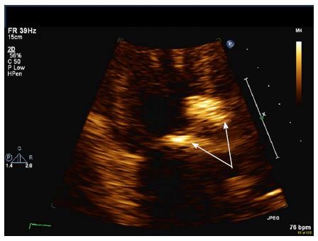
Figure 1 Severe calcification of proximal aorta and aortic leaflets (arrows) resulting in moderate aortic regurgitation and stenosis.
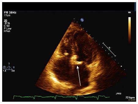
Figure 2 Apical four chamber view of mitral annular calcification (arrow).
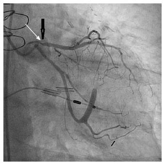
Figure 3 Severe proximal stenosis of the left anterior descending coronary artery (arrow).
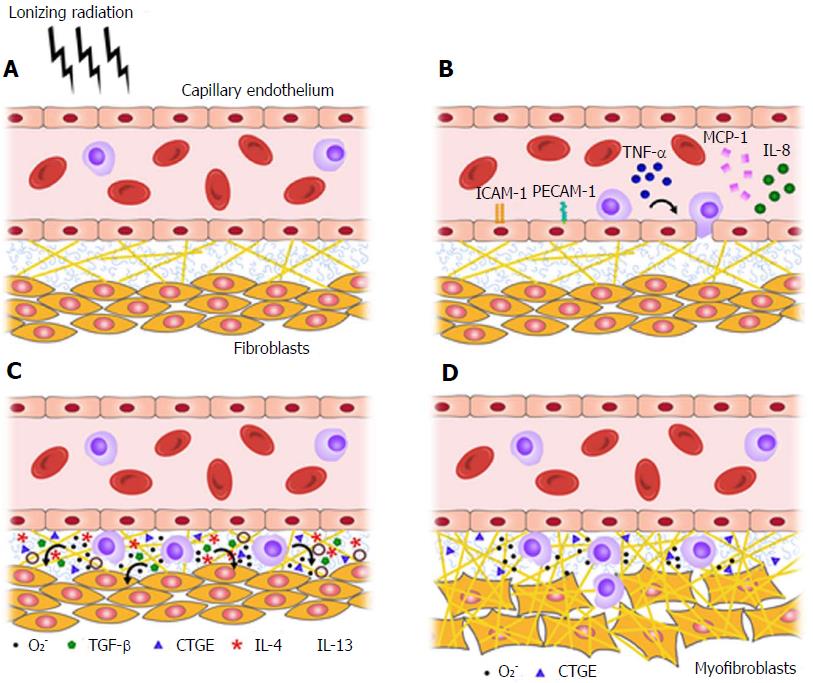
Figure 4 Radiation injury and the transition from acute inflammation to chronic fibrosis, as mediated by pro-fibrotic cytokines and reactive oxygen species.
A-C: Normal tissue (A) becomes inflamed within hours of irradiation (B), and a pro-fibrotic cytokine profile predominates within days-to-weeks (C); D: Represents the chronic state of fibrosis characteristic of radiation injury. O2-: Reactive oxygen species; TNF-α: Tumor necrosis factor alpha; MCP-1: Monocyte chemotactic protein-1; CTGF: Connective tissue growth factor; TGF-β: Tumor growth factor beta; IL: Interleukin.
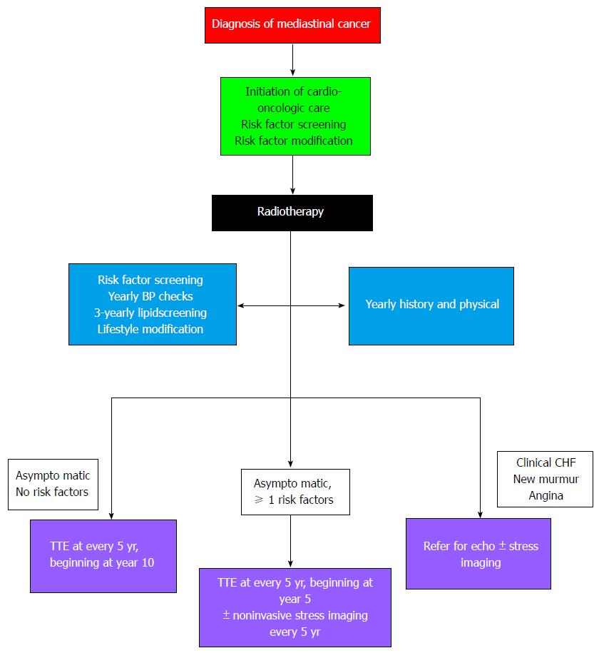
Figure 5 Proposed algorithm for cardio-oncologic screening following mediastinal radiotherapy[84].
CHF: Congestive heart failure; TTE: Transthoracic echocardiography.
- Citation: Cuomo JR, Sharma GK, Conger PD, Weintraub NL. Novel concepts in radiation-induced cardiovascular disease. World J Cardiol 2016; 8(9): 504-519
- URL: https://www.wjgnet.com/1949-8462/full/v8/i9/504.htm
- DOI: https://dx.doi.org/10.4330/wjc.v8.i9.504


