Copyright
©The Author(s) 2016.
World J Cardiol. Aug 26, 2016; 8(8): 488-495
Published online Aug 26, 2016. doi: 10.4330/wjc.v8.i8.488
Published online Aug 26, 2016. doi: 10.4330/wjc.v8.i8.488
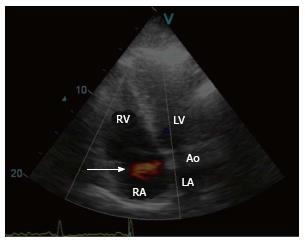
Figure 1 A frame of colored Doppler trans-thoracic echocardiography, five-chamber view illustrating the aortic-atrial fistula (arrow).
Ao: Aorta; RA: Right atrium; LA: Left atrium; RV: Right ventricle; LV: Left ventricle.
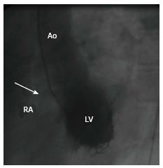
Figure 2 Levo-ventriculogram in the left anterior oblique view demonstrating the fistula (arrow) between the right and non-coronary sinuses communicating with the right atrium.
RCS/NCS: Right and non-coronary sinuses; LV: Levo-ventriculogram; RA: Right atrium.
- Citation: Said SAM, Mariani MA. Acquired aortocameral fistula occurring late after infective endocarditis: An emblematic case and review of 38 reported cases. World J Cardiol 2016; 8(8): 488-495
- URL: https://www.wjgnet.com/1949-8462/full/v8/i8/488.htm
- DOI: https://dx.doi.org/10.4330/wjc.v8.i8.488


