Copyright
©The Author(s) 2016.
World J Cardiol. Jul 26, 2016; 8(7): 401-412
Published online Jul 26, 2016. doi: 10.4330/wjc.v8.i7.401
Published online Jul 26, 2016. doi: 10.4330/wjc.v8.i7.401
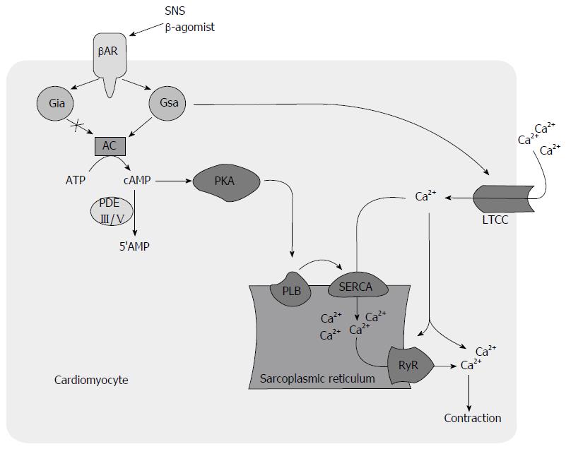
Figure 1 Beta-adrenoreceptor mediated signal transduction leads to the activation of both G stimulatory alpha protein and G inhibitory alpha protein.
Activated Gαs activates adenylyl cyclase (AC) which converts ATP into cAMP while activated Gαi inhibits AC. Activated Gαs also leads to calcium (Ca2+) mobilization into cardiomyocyte by activating L-type calcium channel (LTCC) independent of AC. This increase in intracellular Ca2+ concentration leads to activation of ryanodine receptor (RyR) which causes further release of Ca2+ from SR, a phenomenon known as calcium-induced calcium release. Elevated cAMP activates phosphokinase A (PKA) that inhibits phospholamban (PLB) by phosphorylating it. Phosphorylation of PLB increases uptake of Ca2+ from cytosol into the SR through sarcoplasmic reticulum calcium ATPase (SERCA). This enhanced Ca2+ entry into SR has positive impact on both systolic and diastolic function. In diastole, decreased intracellular Ca2+ causes relaxation. In systole increased release of Ca2+ from SR store through RyR activation increases inotropy. In the failing myocardium, chronic stimulation of βAR results in ineffective activation of AC, persistent activation of L-type calcium channel that increases Ca2+ influx, and decreased Ca2+ uptake into the SR due to decreased SERCA activity. This translates into systolic and diastolic dysfunction and increased arrhythmogenicity. βAR: Beta-adrenoreceptor; ATP: Adenosine triphosphate; cAMP: Cyclic adenosine monophosphate; Gαi: G inhibitory alpha protein; Gαs: G stimulatory alpha protein; PDE: Phosphodiesterase; SNS: Sympathetic nervous system.
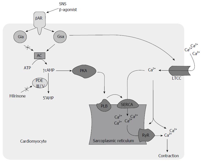
Figure 2 Milrinone causes inhibition of phosphodiesterase III enzyme which decreases cyclic adenosine monophosphate concentration by converting later into inactive 5’adenosine monophosphate.
Increased cyclic adenosine monophosphate (cAMP) activates phosphokinase A (PKA) that inhibits phospholamban (PLB) by phosphorylating it. Inhibition of PLB increases uptake of calcium (Ca2+) from cytosol into the SR through sarcoplasmic reticulum calcium ATPase (SERCA). This enhanced Ca2+ entry into SR has positive impact on both systolic and diastolic function. During diastole, decreased cytosolic Ca2+ causes relaxation. During systole increased release of Ca2+ from SR store through ryanodine receptor (RyR) activation increases inotropy. However, unchecked chronic stimulation of beta-adrenoreceptor (βAR) causes inhibition of AC through Gαi protein and increases intracellular Ca2+ influx by activation of L-type calcium channel (LTCC). Activated LTCC indirectly increases intracellular Ca2+ through activation of RyR mediated release of Ca2+ from SR. This increased intracellular influx of Ca2+ is associated with increased arrhythmogenicity. ATP: Adenosine triphosphate; Gαi: G inhibitory alpha protein; Gαs: G stimulatory alpha protein; PDE: Phosphodiesterase; SNS: Sympathetic nervous system.
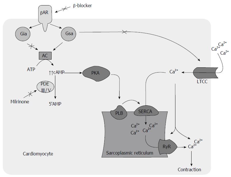
Figure 3 Concomitant use of beta blocker and milrinone causes inhibition of G inhibitory alpha protein which is an inhibitor of adenylyl cyclase and phosphodiesterase III enzyme, both results in increased cyclic adenosine monophosphate concentration.
Increased cAMP inhibits phospholamban (PLB) resulting in efficient movement of calcium (Ca2+) from cytosol into the SR through sarcoplasmic reticulum calcium ATPase (SERCA). This PLB mediated Ca2+ handling results in improved systolic and diastolic function. In addition, BB inhibits beta-adrenoreceptor (βAR) mediated increased Ca2+ influx through L-type calcium channel (LTCC) that is associated with increased arrhythmogenicity. ATP: Adenosine triphosphate; cAMP: Cyclic adenosine monophosphate; Gαi: G inhibitory alpha protein; Gαs: G stimulatory alpha protein; PDE: Phosphodiesterase; SNS: Sympathetic nervous system; BB: Beta blocker; AC: Adenylyl cyclase.
- Citation: Jaiswal A, Nguyen VQ, Le Jemtel TH, Ferdinand KC. Novel role of phosphodiesterase inhibitors in the management of end-stage heart failure. World J Cardiol 2016; 8(7): 401-412
- URL: https://www.wjgnet.com/1949-8462/full/v8/i7/401.htm
- DOI: https://dx.doi.org/10.4330/wjc.v8.i7.401


