Copyright
©The Author(s) 2016.
World J Cardiol. Oct 26, 2016; 8(10): 590-595
Published online Oct 26, 2016. doi: 10.4330/wjc.v8.i10.590
Published online Oct 26, 2016. doi: 10.4330/wjc.v8.i10.590
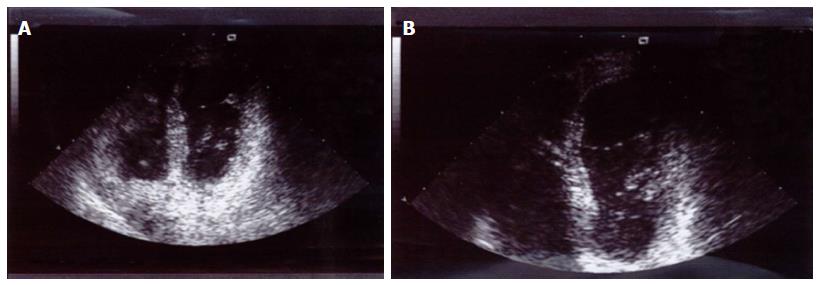
Figure 1 Left ventricular false tendon between the middle segments of the inferior septum and the lateral wall during the cardiac stolic (A) and diastolic (B) cycle.
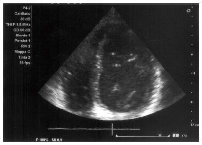
Figure 2 Double false tendon stretched between the lateral wall and inferior septum.
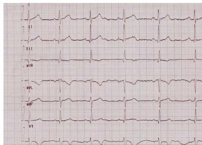
Figure 3 Ventricular repolarization anomalies in precordial leads V1-V4: Inverted T waves from V1 to V3 and flat T waves in D3.
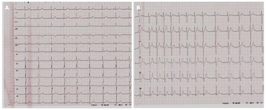
Figure 4 Ventricular repolarization anomalies at rest (A) and normalization after the incremental maximal exercise test on a cycle ergometer (B).
- Citation: Lazarevic Z, Ciminelli E, Quaranta F, Sperandii F, Guerra E, Pigozzi F, Borrione P. Left ventricular false tendons and electrocardiogram repolarization abnormalities in healthy young subjects. World J Cardiol 2016; 8(10): 590-595
- URL: https://www.wjgnet.com/1949-8462/full/v8/i10/590.htm
- DOI: https://dx.doi.org/10.4330/wjc.v8.i10.590


