Copyright
©The Author(s) 2016.
World J Cardiol. Oct 26, 2016; 8(10): 566-574
Published online Oct 26, 2016. doi: 10.4330/wjc.v8.i10.566
Published online Oct 26, 2016. doi: 10.4330/wjc.v8.i10.566
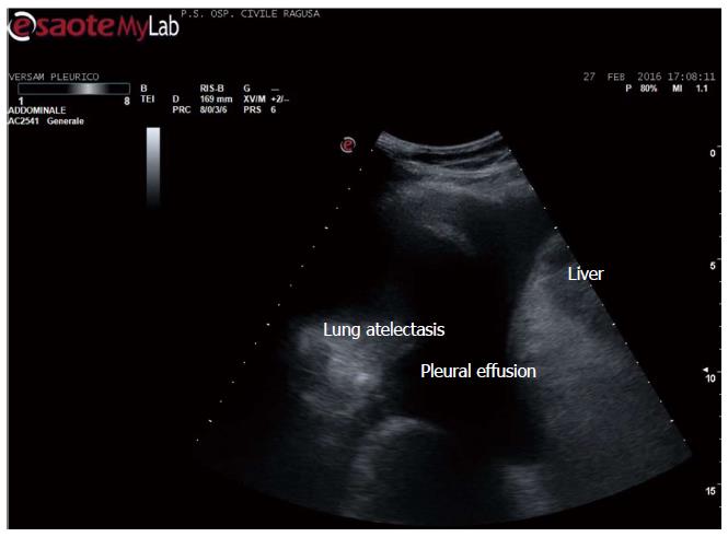
Figure 1 Pleural effusion.
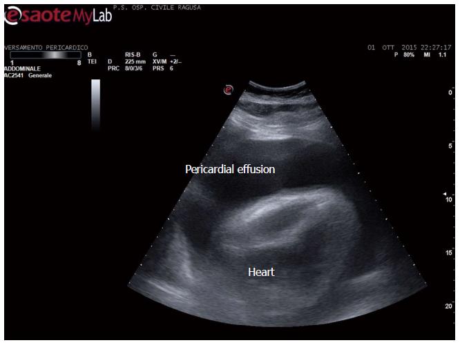
Figure 2 Pericardial and pleural effusion.
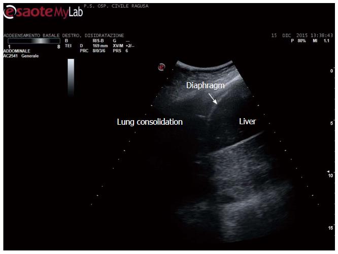
Figure 3 Lung consolidation.
Community acquired pneumonia in an adult.
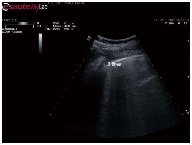
Figure 4 B-lines in acute heart failure.
B-lines count is a dynamic observation, essentially qualitative, since the number changes continuously - from 3 to 6 or more - in case of numerous b-lines. Identical artefacts are detectable in other conditions, including pulmonary fibrosis and dyspnoea due to other causes, including BPCO.
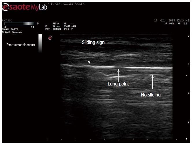
Figure 5 Disappearance of pleural sliding, better demonstrated by video.
Which is here showed as a drop in the continuity of the line, not moving side by side (by courtesy of Giuseppe Molino, MD, MCAU Ospedale Civile di Ragusa, Italy).
- Citation: Trovato GM. Thoracic ultrasound: A complementary diagnostic tool in cardiology. World J Cardiol 2016; 8(10): 566-574
- URL: https://www.wjgnet.com/1949-8462/full/v8/i10/566.htm
- DOI: https://dx.doi.org/10.4330/wjc.v8.i10.566


