Copyright
©The Author(s) 2015.
World J Cardiol. Sep 26, 2015; 7(9): 579-582
Published online Sep 26, 2015. doi: 10.4330/wjc.v7.i9.579
Published online Sep 26, 2015. doi: 10.4330/wjc.v7.i9.579
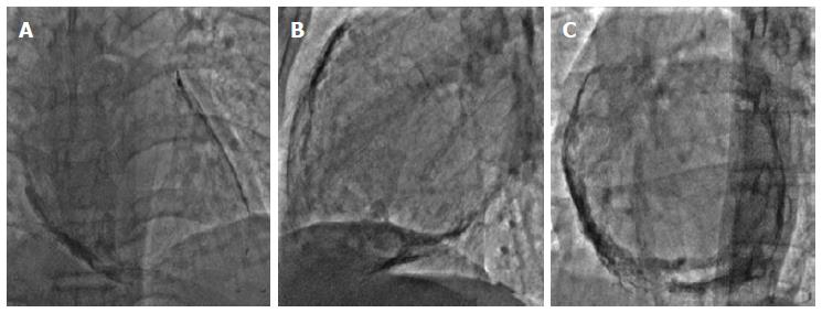
Figure 1 Fluoroscopy images in antero-posterior (A), lateral (B) and left anterior oblique 30º (C) views show circumferential pericardial calcification around the heart.
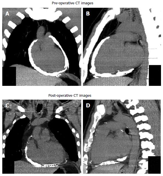
Figure 2 Computed tomography.
Non-contrast computed tomography reconstructed images in coronal (A) and sagittal (B) planes show thick, calcified pericardium around the heart. A repeat CT following partial pericardiectomy in coronal (C) and sagittal (D) planes show residual calcified pericardium at right atrial and posterior surface of the heart. Calcified pericardium is absent along the antero-lateral surface of the heart. CT: Computed tomography.
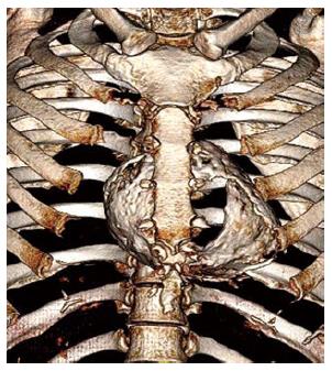
Figure 3 Pre-operative non-contrast computed tomography reconstructed volume rendered image shows pericardial “eggshell” calcification around the heart.
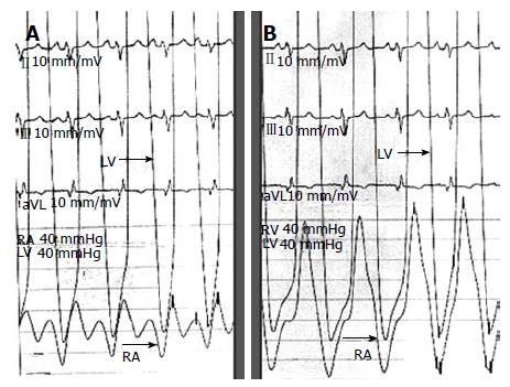
Figure 4 Hemodynamic tracing during catheterization.
A: Right atrial (RA) pressure tracing shows prominent X and Y descent; B: Ventricular pressure tracing shows typical “dip-and-plateau configuration” during diastole. RV: Right ventricle; LV: Left ventricle.
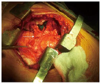
Figure 5 Surgical image shows resected thick and calcified pericardium (white arrow) from anterior surface of heart.
- Citation: Vijayvergiya R, Vadivelu R, Mahajan S, Rana SS, Singhal M. Eggshell calcification of the heart in constrictive pericarditis. World J Cardiol 2015; 7(9): 579-582
- URL: https://www.wjgnet.com/1949-8462/full/v7/i9/579.htm
- DOI: https://dx.doi.org/10.4330/wjc.v7.i9.579


