Copyright
©The Author(s) 2015.
World J Cardiol. Aug 26, 2015; 7(8): 454-465
Published online Aug 26, 2015. doi: 10.4330/wjc.v7.i8.454
Published online Aug 26, 2015. doi: 10.4330/wjc.v7.i8.454
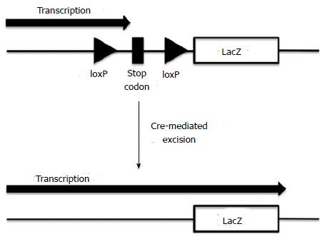
Figure 1 Schematic representation of LacZ expression following the excision of a floxed stop codon by Cre recombinase.
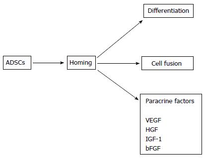
Figure 2 Possible mechanisms underlying the effect of adipose tissue-derived stem cells on regeneration of the cardiovascular system.
ADSCs: Adipose tissue-derived stem cells; VEGF: Vascular endothelial growth factor; HGF: Hepatocyte growth factor; IGF-1: Insulin-like growth factor-1; bFGF: Basic fibroblast growth factor.
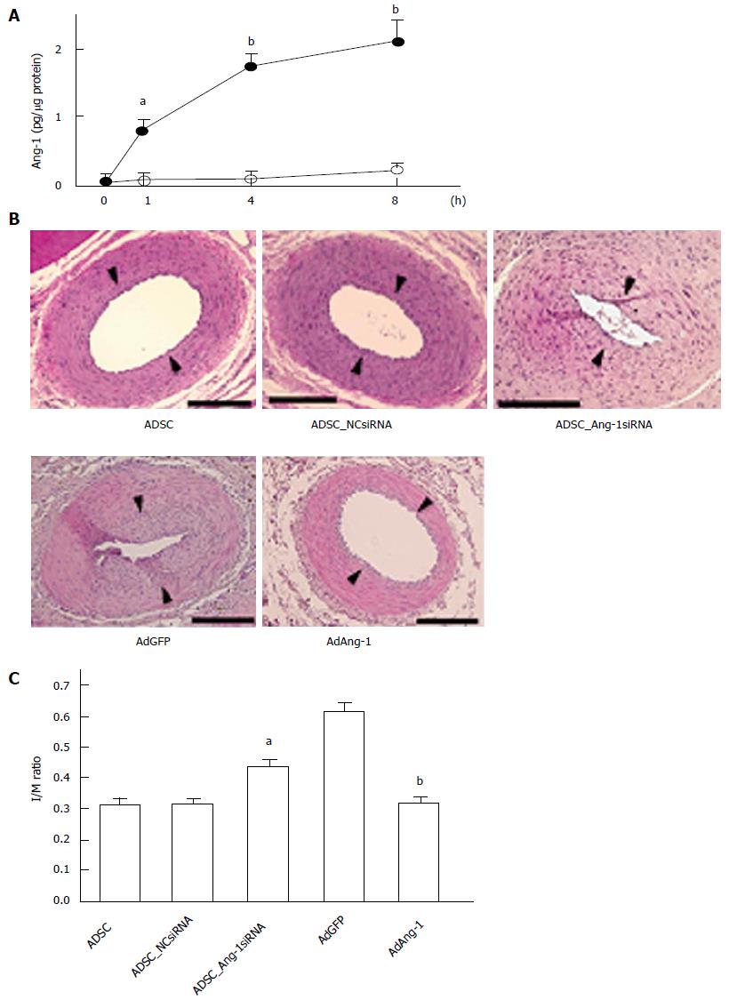
Figure 3 Ang-1 is implicated in Adipose tissue-derived stem cell-induced suppression of neointimal formation.
A: ADSCs produce Ang-1, particularly when cultured in medium containing growth factors for VECs. Rat ADSCs were plated in 24-well plates and cultured in control medium (open circles) or medium containing growth factors for VECs (EGM: closed circles) for 1 wk. After washing with PBS, the medium was replaced with serum-free Dulbecco’s modified Eagle medium and incubated for the indicated periods. Ang-1 accumulation was measured with an enzyme-linked immunosorbent assay kit. aP < 0.05, bP < 0.01 vs 0 h (n = 6 per group); B: Effect of knockdown of endogenous Ang-1 in ADSCs and forced expression of Ang-1 on neointimal formation. ADSCs were infected with lentivirus expressing negative control siRNA (NCsiRNA), which does not suppress the expression of mammalian mRNA, or lentivirus expressing Ang-1 siRNA (Ang-1siRNA). ADSCs not infected with lentivirus were used as positive controls (ADSC). ADSCs were cultured in EGM for 1 wk. ADSCs (106 cells) were seeded from the adventitial side immediately after wire injury of the rat femoral artery. Adenoviruses expressing green fluorescent protein (AdGFP) or Ang-1 (AdAng-1) were also injected into the femoral artery from the adventitial side following wire injury. Femoral arteries were harvested 14 d after injury for histological analyses. Arrowheads indicate the position of internal elastic lamina. Bars represent 100 μm; C: I/M ratios were compared among the groups (n = 8 per group). aP < 0.05 vs NCsiRNA infection and bP < 0.01 vs AdGFP infection. PBS: Phosphate-buffered saline; ADSCs: Adipose tissue-derived stem cells; VECs: Vascular endothelial cells.
- Citation: Suzuki E, Fujita D, Takahashi M, Oba S, Nishimatsu H. Adipose tissue-derived stem cells as a therapeutic tool for cardiovascular disease. World J Cardiol 2015; 7(8): 454-465
- URL: https://www.wjgnet.com/1949-8462/full/v7/i8/454.htm
- DOI: https://dx.doi.org/10.4330/wjc.v7.i8.454


