Copyright
©The Author(s) 2015.
World J Cardiol. Jul 26, 2015; 7(7): 431-433
Published online Jul 26, 2015. doi: 10.4330/wjc.v7.i7.431
Published online Jul 26, 2015. doi: 10.4330/wjc.v7.i7.431
Figure 1 Transthoracic echocardiogram using apical three chamber view showing the big anterior left ventricular aneurysm (arrow).
The wall of the aneurysm was calcified (arrowheads), and the aneurysm was covered with thrombus (arrow). LA: Left atrium; LV: Left ventricle.
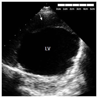
Figure 2 Transthoracic echocardiogram using parasternal short axis view at the midventricular level showing the thrombus (arrow) covering the anterior wall aneurysm.
LV: Left ventricle.
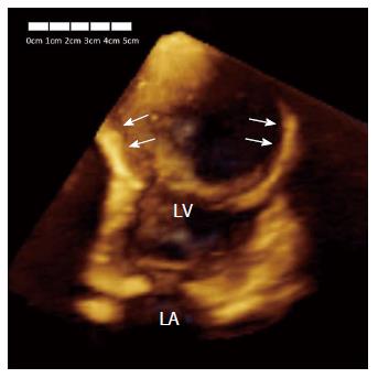
Figure 3 Three-dimensional echocardiography in apical four chamber view showing the big size of the aneurysm (arrows).
LA: Left atrium; LV: Left ventricle.
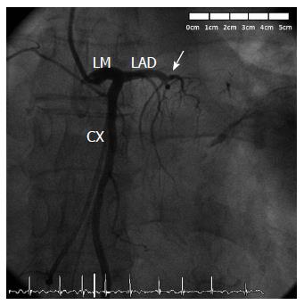
Figure 4 Left coronary angiography demonstrating a proximal occlusion of the left anterior descending artery (arrow).
CX: Circumflex coronary artery; LAD: Left anterior descending coronary; LM: Left main coronary artery.
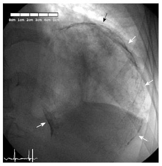
Figure 5 Fluoroscopic imaging in right anterior oblique projection showing a complete oval calcified mass (arrows), corresponding with the left ventricular aneurysm.
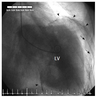
Figure 6 Left ventriculogram confirming diagnosis of a giant calcified and partially thrombosed left ventricular aneurysm, with severe left ventricular systolic dysfunction.
The wall of the aneurysm is calcified (arrowheads), and the aneurysm is covered with thrombus (arrows). LV: Left ventricle.
- Citation: de Agustin JA, de Diego JJG, Marcos-Alberca P, Rodrigo JL, Almeria C, Mahia P, Luaces M, Garcia-Fernandez MA, Macaya C, de Isla LP. Giant and thrombosed left ventricular aneurysm. World J Cardiol 2015; 7(7): 431-433
- URL: https://www.wjgnet.com/1949-8462/full/v7/i7/431.htm
- DOI: https://dx.doi.org/10.4330/wjc.v7.i7.431


