Copyright
©The Author(s) 2015.
World J Cardiol. Jun 26, 2015; 7(6): 367-372
Published online Jun 26, 2015. doi: 10.4330/wjc.v7.i6.367
Published online Jun 26, 2015. doi: 10.4330/wjc.v7.i6.367
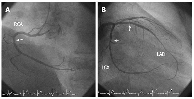
Figure 1 Coronary angiography before spasm provocation tests.
A: There were mild atherosclerotic changes at the proximal segment of the right coronary artery (RCA); B: There were mild atherosclerotic changes at the proximal segment of the left anterior descending coronary artery (LAD) and at the mid-segment of the left circumflex coronary artery (LCX). The mild atherosclerotic changes are indicated using arrows.
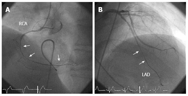
Figure 2 Coronary angiography during the spasm provocation tests.
A: In the right coronary artery (RCA), a diffuse coronary spasm occurred at the mid-distal segments after the intracoronary infusion of 50 μg acetylcholine (ACh); B: In the left coronary artery, a diffuse coronary spasm occurred at the mid-distal segments of the left anterior descending coronary artery (LAD) after the intracoronary infusion of 100 μg ACh. The coronary spasm segments are indicated using arrows.
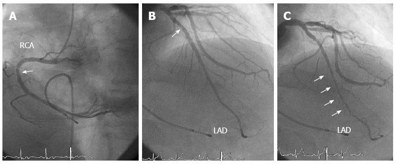
Figure 3 Coronary angiography after the intracoronary infusion of nitroglycerin.
A: There was a mild atherosclerotic change at the proximal segment of the right coronary artery (RCA); B: There was a mild atherosclerotic change (indicated with arrows) in the proximal segment of the left anterior descending coronary artery (LAD) at the end-diastolic phase; C: There was myocardial bridging (indicated with arrows) at the mid-distal segments of the LAD at the end-systolic phase.
Figure 4 The fractional flow reserve using the intravenous infusion of adenosine triphosphate was 0.
77 (A), which jumped up to 0.90 through the myocardial bridging during pullback (B).
- Citation: Teragawa H, Fujii Y, Ueda T, Murata D, Nomura S. Case of angina pectoris at rest and during effort due to coronary spasm and myocardial bridging. World J Cardiol 2015; 7(6): 367-372
- URL: https://www.wjgnet.com/1949-8462/full/v7/i6/367.htm
- DOI: https://dx.doi.org/10.4330/wjc.v7.i6.367


