Copyright
©The Author(s) 2015.
World J Cardiol. Jun 26, 2015; 7(6): 357-360
Published online Jun 26, 2015. doi: 10.4330/wjc.v7.i6.357
Published online Jun 26, 2015. doi: 10.4330/wjc.v7.i6.357
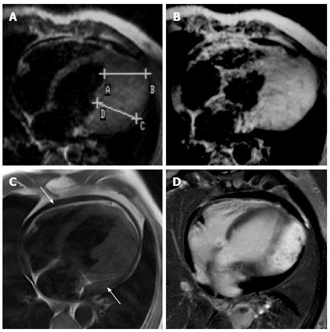
Figure 1 Hypertrophic cardiomyopathy of the left lateral wall in 1989 (A, B) and 2012 (C, D).
Note the difference of image quality especially within the GE series from 1989 (B) and the PSIR-SSFP late Gadolinium enhancement (LGE) images in 2012 (D). Within the last 23 years, there was no significant change in respect to morphology (A, C) and LGE (B, D) in this patient. Arrows indicate accompanying circumferential, increasing pericardial effusion.
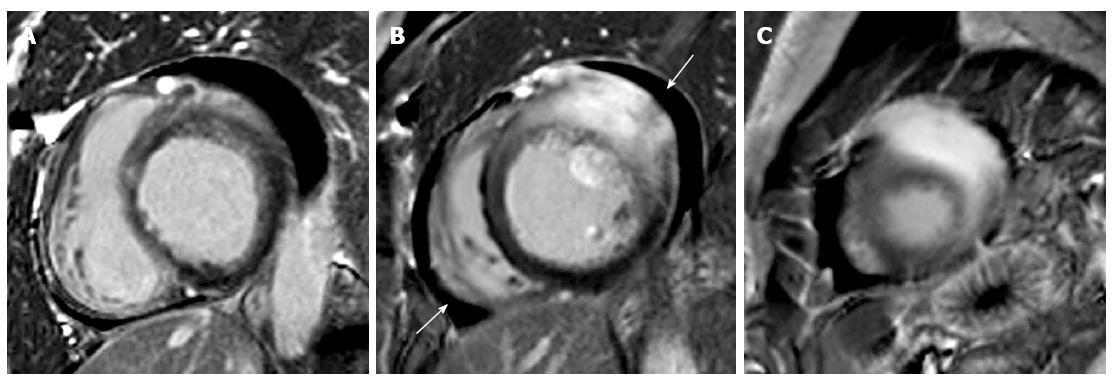
Figure 2 Late Gadolinium enhancement images in basal (A), midventricular (B) and apical (C) slices in the short axis view show significant late Gadolinium enhancement in the area of hypertrophic cardiomyopathy of the left lateral wall.
Arrows indicate accompanying pericardial effusion.
- Citation: Gassenmaier T, Petritsch B, Kunz AS, Gkaniatsas S, Gaudron PD, Weidemann F, Nordbeck P, Beer M. Long term evolution of magnetic resonance imaging characteristics in a case of atypical left lateral wall hypertrophic cardiomyopathy. World J Cardiol 2015; 7(6): 357-360
- URL: https://www.wjgnet.com/1949-8462/full/v7/i6/357.htm
- DOI: https://dx.doi.org/10.4330/wjc.v7.i6.357


