Copyright
©The Author(s) 2015.
World J Cardiol. May 26, 2015; 7(5): 293-298
Published online May 26, 2015. doi: 10.4330/wjc.v7.i5.293
Published online May 26, 2015. doi: 10.4330/wjc.v7.i5.293
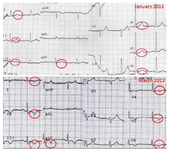
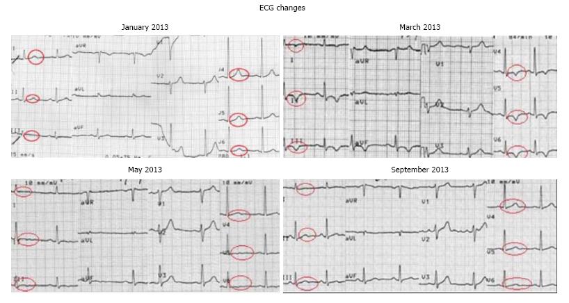
Figure 1 Electrocardiogram changes: Ventricular.
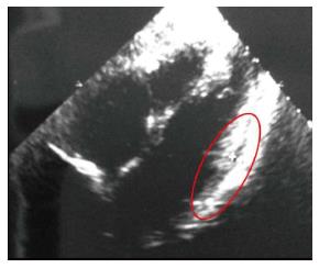
Figure 2 Echocardiogram (March 2013): Echo-free space in the lateral wall and hyper echogenicity of the lateral portion of the left ventricle.
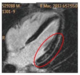
Figure 3 Cardiac magnetic resonance: Areas of intramural and sub-epicardial delayed enhancement in the lateral wall, suggestive of fibrosis.
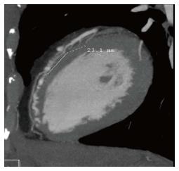
Figure 4 Tomographic computed scan: Myocardial bridge (23 mm).
Figure 5 Electrocardiogram follow up.
- Citation: Quaranta F, Guerra E, Sperandii F, Santis FD, Pigozzi F, Calò L, Borrione P. Myocarditis in athlete and myocardial bridge: An innocent bystander? World J Cardiol 2015; 7(5): 293-298
- URL: https://www.wjgnet.com/1949-8462/full/v7/i5/293.htm
- DOI: https://dx.doi.org/10.4330/wjc.v7.i5.293


