Copyright
©The Author(s) 2015.
World J Cardiol. May 26, 2015; 7(5): 230-237
Published online May 26, 2015. doi: 10.4330/wjc.v7.i5.230
Published online May 26, 2015. doi: 10.4330/wjc.v7.i5.230
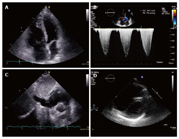
Figure 1 Characteristic echocardiogram of a patient with pulmonary hypertension.
A: Apical 4-chamber view showing dilatation of the right ventricle; B: Apical 4-chamber view with Doppler signal (continuous wave) showing an increased right ventricular- right atrial pressure gradient (4.4 m/s); C: Subcostal view showing dilatation of the inferior caval vein corresponding with an increased pressure in the right atrium; D: Parasternal short axis view showing flattening of the interventricular septum (D-sign) and dilatation of the right ventricle.
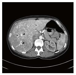
Figure 2 Hepatic arteriovenous malformations.
Computed tomography with contrast in arterial fase showing extensive filling of the hepatic veins (arrows), and diffuse hepatic arteriovenous malformations (asterix).
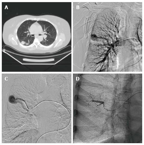
Figure 3 Pulmonary arteriovenous malformations.
A: Computed tomography of the chest with large pulmonary arteriovenous malformation (PAVM) in the lower lobe of the right lung; B: Pulmonary angiogram of the PAVM in the same patient; C: Selective pulmonary angiogram of the PAVM in the apex of the lower lobe of the right lung; D: Repeat angiogram after transcatheter embolisation of the PAVM with a vascular plug.
- Citation: Vorselaars VM, Velthuis S, Snijder RJ, Vos JA, Mager JJ, Post MC. Pulmonary hypertension in hereditary haemorrhagic telangiectasia. World J Cardiol 2015; 7(5): 230-237
- URL: https://www.wjgnet.com/1949-8462/full/v7/i5/230.htm
- DOI: https://dx.doi.org/10.4330/wjc.v7.i5.230


