Copyright
©The Author(s) 2015.
World J Cardiol. Apr 26, 2015; 7(4): 167-177
Published online Apr 26, 2015. doi: 10.4330/wjc.v7.i4.167
Published online Apr 26, 2015. doi: 10.4330/wjc.v7.i4.167
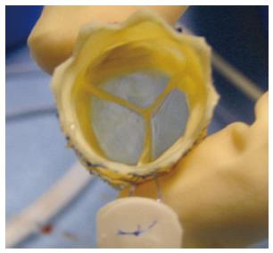
Figure 1 The MELODYTM percutaneous pulmonary valve (Medtronic, MN).
The Melody device in “en face” view. Note the blue outflow line to identify the outflow end of the device (courtesy of P.Lurz).
Figure 2 MELODYTM device and its delivery system.
A: Uncrimped device on the delivery system with a retractable sheath; B: Crimped device before covering the device to protect it during the delivery; C: Crimped and covered device prepared for delivery (courtesy of Lurz P).
Figure 3 The Edwards SAPIENTM pulmonic transcatheter heart valve (Edwards Lifesciences LLC, Irvine, CA).
The SAPIENTM device in lateral view mounted on its delivery system RetroflexTM III (courtesy of Edwards Lifesciences LLC).
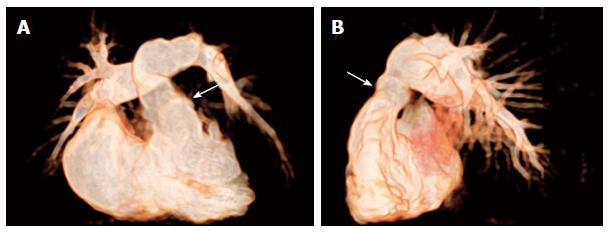
Figure 4 Non-invasive 3D whole heart imaging by magnetic resonance tomography.
Non-invasive 3D whole heart imaging by magnetic resonance tomography was performed in a patient with pulmonary atresia with intact ventricular septum after repair by pulmonic homograft implantation (arrows) with RVOT dysfunction prior to PPVI (A) in a.p. and (B) in lat. view. (Courtesy of Wagner R). RVOT: Right ventricular outflow tract; PPVI: Percutaneous pulmonary valve implantation.
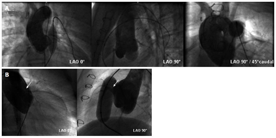
Figure 5 Assessment of risk for coronary compression.
A: Aortic root angiogram and simultaneous (high-pressure) balloon inflation within the eligible landing zone is performed to rule out potential for coronary compression (courtesy of Wagner R); B: Aortic root angiogram showing compression of the left anterior descending coronary artery (arrows) during balloon inflation in the conduit. The procedure was therefore abandoned in this patient and no percutaneus pulmonary valve implantation was performed (courtesy of Wagner R).
Figure 6 MELODY™ device for “Valve-in-Valve” implantation in tricuspid position.
MELODY™ valve implantation procedure in a patient with severe stenosis of a prosthetic biological tricuspid valve (arrow): fluroscopy reveals (A) a RV angiogram in systoly prior to implantation (B) guidewire in the right PA and stable device position after inflating of both balloons of the BiB-delivery system and (C) RV angiogram showing stent position within the biological prosthesis and relative to the ventricle. There is no tricuspid regurgitation. (Courtesy of Wagner R). RV: Right ventricular; PA: Pulmonary artery.
- Citation: Wagner R, Daehnert I, Lurz P. Percutaneous pulmonary and tricuspid valve implantations: An update. World J Cardiol 2015; 7(4): 167-177
- URL: https://www.wjgnet.com/1949-8462/full/v7/i4/167.htm
- DOI: https://dx.doi.org/10.4330/wjc.v7.i4.167


