Copyright
©The Author(s) 2015.
World J Cardiol. Feb 26, 2015; 7(2): 104-110
Published online Feb 26, 2015. doi: 10.4330/wjc.v7.i2.104
Published online Feb 26, 2015. doi: 10.4330/wjc.v7.i2.104
Figure 1 Electrocardiogram on admission.
Electrocardiogram shows an idioventricular rhythm with wide QRS complexes. A clear ST elevation is shown in lead aVR.
Figure 2 Urgent coronary angiography.
A coronary angiography of RCA reveals no stenosis (A), but there is severe stenosis from the ostium of the left main coronary artery to the LAD with TIMI grade 1 flow (B). RCA: Right coronary artery; LMCA: Left main coronary artery; LAD: Left anterior descending artery; TIMI: Thrombolysis in myocardial infarction.
Figure 3 Coronary angiography and intravascular ultrasound findings prior to stent implantation.
IVUS shows compression of the true lumen from the LMCA ostium to the LAD by a large false lumen. A: The large hematoma (arrowhead) eviscerates into the aorta (Ao); B: The false lumen (arrowhead) compresses the LMCA (LM), including both the LAD and LCX ostium. The arrow indicates the guidewire in the LCX; C: The large false lumen (arrowhead) compresses the true lumen (T) in the proximal LAD; D: The false lumen disappears before the bifurcation of the diagonal branch. Scale measures in 1 mm. IVUS: Intravascular ultrasound; LCX: Left circumflex artery; LAD: Left anterior descending artery.
Figure 4 Coronary angiography and intravascular ultrasound findings after stent implantation.
The implanted stent appears to be well expanded and completely seals the false lumen. There was no evidence of distal propagation of the false lumen or a flow limitation of the LCX. A: The hematoma (arrowhead) in the aorta is well covered by the stent; B, C: Implanted stent completely sealed the hematoma (arrowhead); D: No extension of the hematoma beyond the stent. Scale bars represent 1 mm.
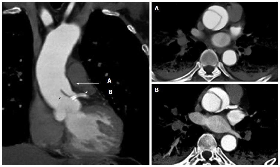
Figure 5 Contrast-enhanced computed tomography after coronary stenting.
The sagittal view (left image) reveals the localized dissection of the ascending aorta that extends to the left main coronary artery ostium. The implanted coronary stent (arrow) protects against the intrusion of the dissection flap (black arrowhead) into the left coronary artery. Ascending aortic dissection is clearly visible in the horizontal view (right images) in accordance with A and B in the sagittal view.
- Citation: Hanaki Y, Yumoto K, I S, Aoki H, Fukuzawa T, Watanabe T, Kato K. Coronary stenting with cardiogenic shock due to acute ascending aortic dissection. World J Cardiol 2015; 7(2): 104-110
- URL: https://www.wjgnet.com/1949-8462/full/v7/i2/104.htm
- DOI: https://dx.doi.org/10.4330/wjc.v7.i2.104


