Copyright
©The Author(s) 2015.
World J Cardiol. Dec 26, 2015; 7(12): 931-937
Published online Dec 26, 2015. doi: 10.4330/wjc.v7.i12.931
Published online Dec 26, 2015. doi: 10.4330/wjc.v7.i12.931
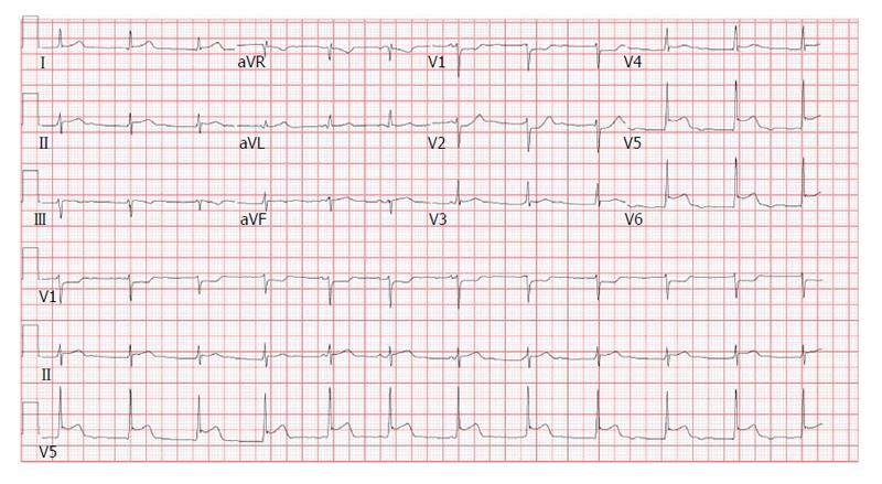
Figure 1 Electrocardiogram on admission with ST segment changes; ST segment depression in V1 and V2 with ST segment elevations in V5 and V6.
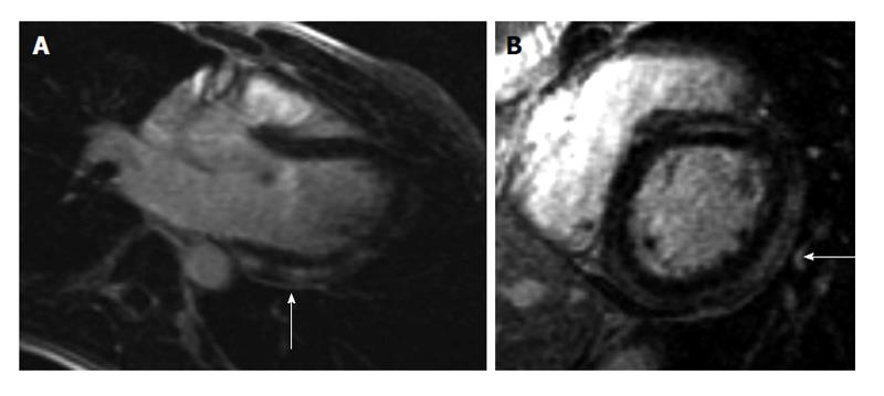
Figure 2 Cardiac magnetic resonance imaging findings.
Cardiac magnetic resonance demonstrates pathological delayed gadolinium enhancement (as indicated by arrows). A: Long axis view: Delayed enhancement of the myocardium demonstrates subepicardial and mid myocardial enhancement indicative of myocarditis; B: Short axis view: Delayed enhancement of the myocardium demonstrates subepicardial and mid myocardial enhancement indicative of myocarditis.
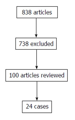
Figure 3 Flow chart of literature review.
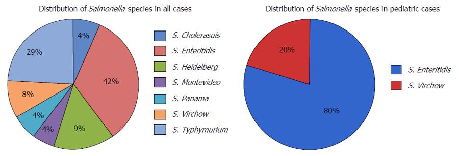
Figure 4 Distribution of Salmonella species in all cases, including pediatric cases.
- Citation: Villablanca P, Mohananey D, Meier G, Yap JE, Chouksey S, Abegunde AT. Salmonella Berta myocarditis: Case report and systematic review of non-typhoid Salmonella myocarditis. World J Cardiol 2015; 7(12): 931-937
- URL: https://www.wjgnet.com/1949-8462/full/v7/i12/931.htm
- DOI: https://dx.doi.org/10.4330/wjc.v7.i12.931


