Copyright
©The Author(s) 2015.
World J Cardiol. Nov 26, 2015; 7(11): 765-775
Published online Nov 26, 2015. doi: 10.4330/wjc.v7.i11.765
Published online Nov 26, 2015. doi: 10.4330/wjc.v7.i11.765
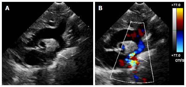
Figure 1 Echocardiogram of coarctation.
A: Two-dimensional transthoracic echocardiogram image obtained from the suprasternal notch in an 11-day-old infant demonstrating discrete coarctation (arrow); B: Color Doppler of the same image with aliasing of flow at the site of coarctation (arrow).
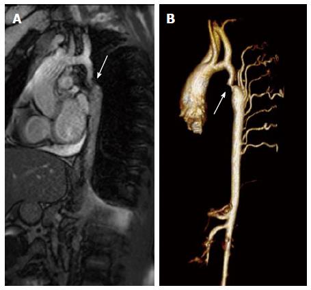
Figure 2 Magnetic resonance imaging of coarctation.
A: Magnetic resonance image (steady-state free precession) in a sagittal projection demonstrating transverse arch hypoplasia and long segment coarctation of the aorta distal to the left subclavian artery (arrow) in a 12-year-old male; B: Three-dimensional reconstruction of a gated contrasted angiogram for the same patient, which demonstrates transverse arch hypoplasia, coarctation at the aorta at the distal transverse aortic arch and isthmus (arrow), and dilated intercostal arteries (collaterals).
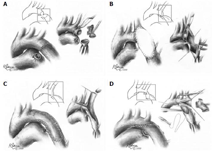
Figure 3 Surgical techniques in coarctation repair.
A: Resection and simple end-to-end anastomosis. The coarctation is resected, and an end-to-end, circumferential anastomosis is created; B: Patch aortoplasty. An incision is extended across the coarctation, and a patch is sutured in place to enlarge the stenotic region; C: Subclavian flap aortoplasty. The left subclavian artery is ligated and divided. A longitudinal incision is extended from the proximal left subclavian artery beyond the area of coarctation, and the proximal left subclavian stump is folded down to enlarge the area of coarctation; D: Resection with extended end-to-end anastomosis. The coarctation is resected using a broad, longitudinal incision, and an oblique anastomosis is constructed between the undersurface of the transverse arch and the descending thoracic aorta. Figures adapted and reprinted with permission from the Journal of Cardiac Surgery[30].
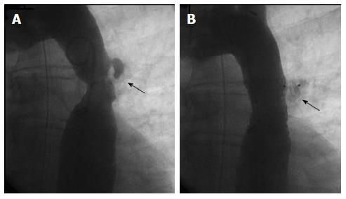
Figure 4 Endovascular stent placement for coarctation.
A: Angiogram (LAO 30°, caudal 30°) demonstrating a discrete coarctation and intercostal aneurysm (arrow) in a 45-year-old male; B: Angiogram in the same projection after endovascular bare metal stent placement showing no significant residual stenosis. The intercostal aneurysm was successfully occluded with an Amplatzer Vascular Plug II (arrow).
- Citation: Torok RD, Campbell MJ, Fleming GA, Hill KD. Coarctation of the aorta: Management from infancy to adulthood. World J Cardiol 2015; 7(11): 765-775
- URL: https://www.wjgnet.com/1949-8462/full/v7/i11/765.htm
- DOI: https://dx.doi.org/10.4330/wjc.v7.i11.765


