Copyright
©The Author(s) 2015.
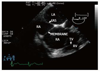
Figure 1 Four-chamber transesophageal echocardiogram view showing the division of the left atrium into two chambers by a transverse membrane.
IAS: Interatrial septum; LA: Left atrium; RA: Right atrium; RV: Right ventricle; TV: Tricuspid valve.
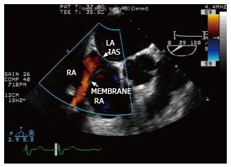
Figure 2 Color flow Doppler demonstrating the partially obstructive nature of the membrane, with blood flow through a small connection in the superior portion of the right atrium between the two chambers.
IAS: Interatrial septum; LA: Left atrium; RA: Right atrium.
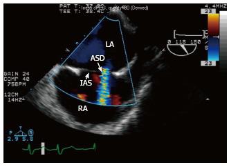
Figure 3 Color flow Doppler demonstrating a small atrial septal defect with left-to-right shunt.
ASD: Atrial septal defect; IAS: Interatrial septum; LA: Left atrium; RA: Right atrium.
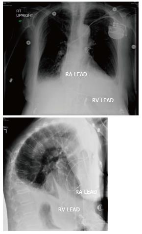
Figure 4 Chest X-ray showing proper placement of right ventricular and right atrial leads.
RA: Right atrium; RV: Right ventricle.
- Citation: Xiang K, Moukarbel GV, Grubb B. Permanent transvenous pacemaker implantation in a patient with Cor triatriatum dextrum. World J Cardiol 2015; 7(1): 43-46
- URL: https://www.wjgnet.com/1949-8462/full/v7/i1/43.htm
- DOI: https://dx.doi.org/10.4330/wjc.v7.i1.43


