Copyright
©2014 Baishideng Publishing Group Inc.
World J Cardiol. Sep 26, 2014; 6(9): 1045-1048
Published online Sep 26, 2014. doi: 10.4330/wjc.v6.i9.1045
Published online Sep 26, 2014. doi: 10.4330/wjc.v6.i9.1045
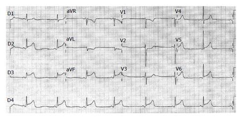
Figure 1 Twelve-lead electrocardiogram showing sinus rhythm with ST-segment elevation in the inferior leads and mirror image (mild ST-segment depression) in V1 to V3 and aVL.
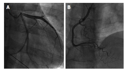
Figure 2 A and B coronary angiography showing angiographically normal coronary arteries.
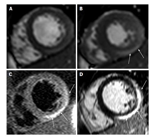
Figure 3 Cardiovascular magnetic resonance.
Upper panel: Still frames of cine movies a t end-diastole (A) and end-systole (B) showing mild infero-lateral hypokinesis (arrows); C: T2-STIR image showing myocardial edema in the lateral and inferior lateral epicardial wall (arrows); D: Gadolinium-enhanced image showing late enhancement predominately in the epicardial lateral and infero-lateral wall (arrows), highly compatible with acute myocarditis.
- Citation: Kumar A, Bagur R, Béliveau P, Potvin JM, Levesque P, Fillion N, Tremblay B, Larose &, Gaudreault V. Acute myocarditis triggering coronary spasm and mimicking acute myocardial infarction. World J Cardiol 2014; 6(9): 1045-1048
- URL: https://www.wjgnet.com/1949-8462/full/v6/i9/1045.htm
- DOI: https://dx.doi.org/10.4330/wjc.v6.i9.1045


