Copyright
©2014 Baishideng Publishing Group Inc.
World J Cardiol. Sep 26, 2014; 6(9): 1038-1040
Published online Sep 26, 2014. doi: 10.4330/wjc.v6.i9.1038
Published online Sep 26, 2014. doi: 10.4330/wjc.v6.i9.1038
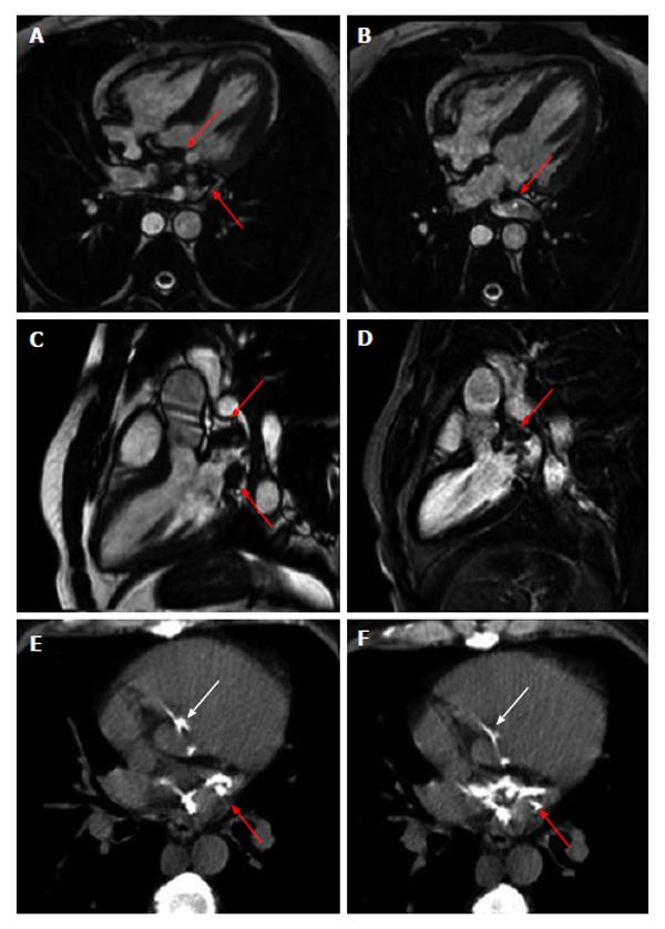
Figure 1 Photograph.
A-D: Cardiac magnetic resonance showed hypointense areas located in left atrium and atrio-ventricular plane (red arrows); B: Partial obstruction of superior pulmonary vein; E and F: Cardiac computed tomography showed the presence of a mass suggestive of calcium in left atrium (red arrows), atrioventricular groove and aortic left ventricular outflow (white arrows).
- Citation: Dattilo G, Anfuso C, Casale M, Giugno V, Camarda L, Laganà N, Di Bella G. Calcific left atrium: A rare consequence of endocarditis. World J Cardiol 2014; 6(9): 1038-1040
- URL: https://www.wjgnet.com/1949-8462/full/v6/i9/1038.htm
- DOI: https://dx.doi.org/10.4330/wjc.v6.i9.1038


