Copyright
©2014 Baishideng Publishing Group Inc.
World J Cardiol. Aug 26, 2014; 6(8): 764-770
Published online Aug 26, 2014. doi: 10.4330/wjc.v6.i8.764
Published online Aug 26, 2014. doi: 10.4330/wjc.v6.i8.764
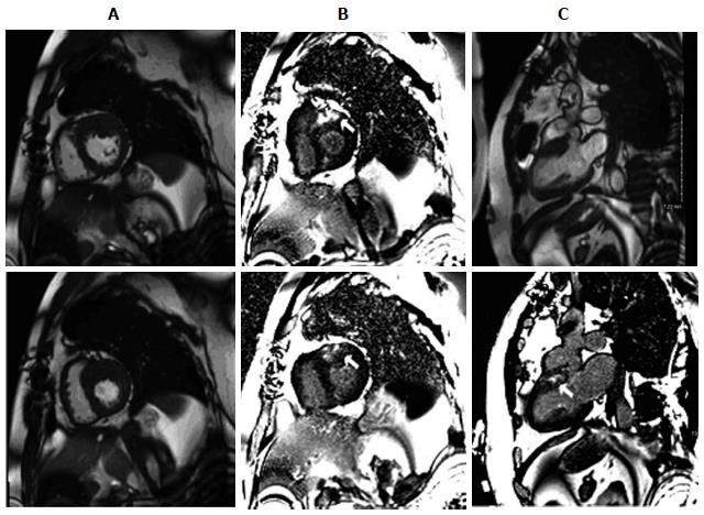
Figure 1 Basal anterior hypertrophic cardiomyopathy in a 45-year-old man with a history of syncope.
Cardiac magnetic resonance imaging demonstrated severe thickening of basal anterior wall with a maximal measurement of 25 mm. A: Two chamber short-axis cine images; B: Late delayed gadolinium enhanced images; C: Two chamber long-axis cine images (top panel). Late delayed gadolinium enhanced images (bottom panel). Patchy, non-coronary artery disease scarring in the hypertrophied areas is indicated by white arrows.
- Citation: Zhang L, Mmagu O, Liu L, Li D, Fan Y, Baranchuk A, Kowey PR. Hypertrophic cardiomyopathy: Can the noninvasive diagnostic testing identify high risk patients? World J Cardiol 2014; 6(8): 764-770
- URL: https://www.wjgnet.com/1949-8462/full/v6/i8/764.htm
- DOI: https://dx.doi.org/10.4330/wjc.v6.i8.764


