Copyright
©2014 Baishideng Publishing Group Inc.
World J Cardiol. Jul 26, 2014; 6(7): 689-691
Published online Jul 26, 2014. doi: 10.4330/wjc.v6.i7.689
Published online Jul 26, 2014. doi: 10.4330/wjc.v6.i7.689
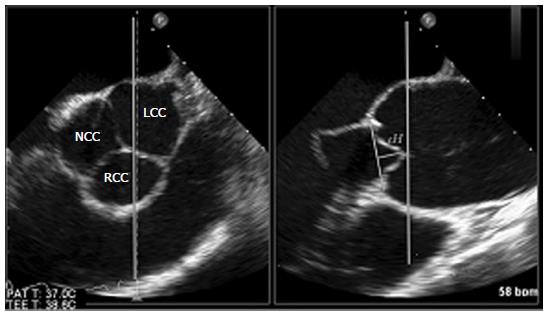
Figure 1 Established method of measuring the effective height in the 2-D short axis (left) and long axis image of the proximal aorta (right).
As shown, only the coaptation between the right coronary cusp anteriorly and the left coronary cusp posteriorly can be measured. RCC: Right coronary cusp; LCC: Left coronary cusp; NCC: Noncoronary cusp.
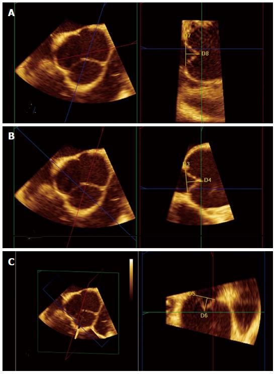
Figure 2 Images of a normal aortic valve from multiple plane reconstruction of the aortic valve with 3D transoesophageal echocardiography.
A: The red plane intersects the coaptation surface of the noncoronary cusp (NCC) and left coronary cusps (LCC) The yellow line labelled D8 (12.7 mm) represents the LCC effective height; B: The red plane intersects the coaptation surface of the LCC and right coronary cusps (RCC) The yellow line labelled D4 (13.9 mm) represents the effective height; C: The red plane intersects the coaptation surface of the NCC and RCC. The yellow line labelled D6 (12.7 mm) represents the effective height.
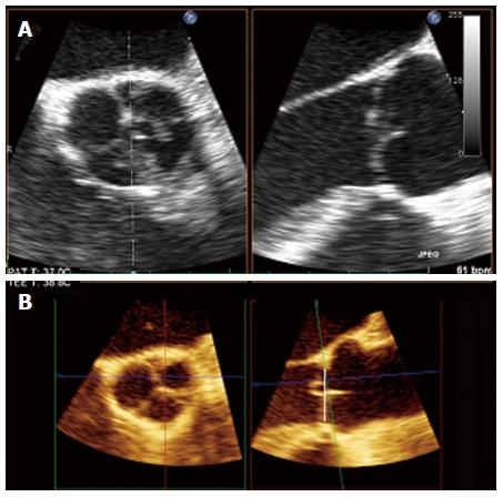
Figure 3 Patient male, 53-year-old.
A: On 2 D there is no clear demonstration of prolapse; B: Multiple plane reconstruction of the aortic valve with 3D transesophageal echocardiography: on reformatted image a prolapse of left coronary cusps is shown (right).
- Citation: Nijs J, Gelsomino S, Kietselaer BB, Parise O, Lucà F, Maessen JG, Meir ML. 3D-echo in preoperative assessment of aortic cusps effective height. World J Cardiol 2014; 6(7): 689-691
- URL: https://www.wjgnet.com/1949-8462/full/v6/i7/689.htm
- DOI: https://dx.doi.org/10.4330/wjc.v6.i7.689


