Copyright
©2014 Baishideng Publishing Group Inc.
World J Cardiol. Jul 26, 2014; 6(7): 685-688
Published online Jul 26, 2014. doi: 10.4330/wjc.v6.i7.685
Published online Jul 26, 2014. doi: 10.4330/wjc.v6.i7.685
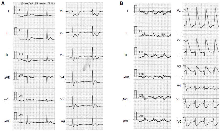
Figure 1 Electrocardiograms showed complete atrioventricular block when blood pressure decreased (A) and ST elevations were noted in leads II, III, aVF, and V1-3 immediately after cardiopulmonary resuscitation (B).
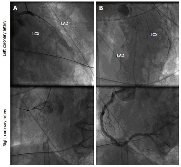
Figure 2 Emergent coronary angiography showed severe narrowing of the left anterior descending coronary artery and the left circumflex coronary artery and occlusion at the proximal segment of the right coronary artery (indicated by an arrow) in initial shots (A), and the trunks of the left anterior descending coronary artery, left circumflex coronary artery, and right coronary artery were found to be dilated despite remaining coronary spasm at the branches after intracoronary infusions of nitroglycerin and epinephrine (B).
LAD: Left anterior descending coronary artery; LCX: Left circumflex coronary artery.
- Citation: Teragawa H, Nishioka K, Fujii Y, Idei N, Hata T, Kurushima S, Shokawa T, Kihara Y. Worsening of coronary spasm during the perioperative period: A case report. World J Cardiol 2014; 6(7): 685-688
- URL: https://www.wjgnet.com/1949-8462/full/v6/i7/685.htm
- DOI: https://dx.doi.org/10.4330/wjc.v6.i7.685


