Copyright
©2014 Baishideng Publishing Group Inc.
World J Cardiol. May 26, 2014; 6(5): 349-352
Published online May 26, 2014. doi: 10.4330/wjc.v6.i5.349
Published online May 26, 2014. doi: 10.4330/wjc.v6.i5.349
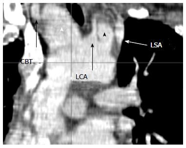
Figure 1 2D Coronal reformatted image.
Digital multiplane reformatted image of the aortic arch, depicting the double barrel-shaped contained rupture of the aortic arch, in-between the common brachial trunk (CBT) and the left carotid artery (LCA) (white arrowhead) and LCA and the left subclavian artery (LSA) (black arrowhead).
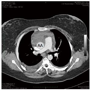
Figure 2 Axial plane computed tomography image of the ascending aorta.
Axial image of the ascending aorta at the level of the pulmonary artery bifurcation. The ascending aorta is compressed in an oval shape due to the sub-adventitial spreading hematoma. AA: Ascending aorta.
- Citation: Nijs J, Gelsomino S, Lucà F, Parise O, Maessen JG, Meir ML. Unreliability of aortic size index to predict risk of aortic dissection in a patient with Turner syndrome. World J Cardiol 2014; 6(5): 349-352
- URL: https://www.wjgnet.com/1949-8462/full/v6/i5/349.htm
- DOI: https://dx.doi.org/10.4330/wjc.v6.i5.349


