Copyright
©2014 Baishideng Publishing Group Inc.
World J Cardiol. May 26, 2014; 6(5): 295-303
Published online May 26, 2014. doi: 10.4330/wjc.v6.i5.295
Published online May 26, 2014. doi: 10.4330/wjc.v6.i5.295
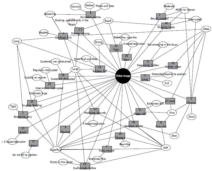
Figure 1 Pulse image network.
The classic pathologic 27 pulse images (greyish, rectangular nodes) described by common attributes (whitish, ellipsoid nodes) derived from categories (frequency, rhythm, wideness, depth, and qualities). Notice that there are pulse images described by exclusive attributes, while other pulse images are described by shared attributes.
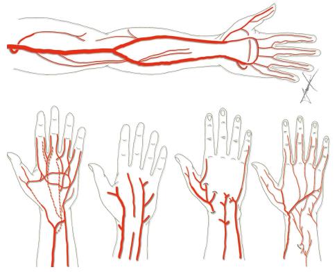
Figure 2 Anatomical drawings on variations of the course of the radial artery.
Top: Most frequent arterial pattern of the radial artery. Bottom: Examples of anatomical variations of the radial artery at the wrist.
- Citation: Ferreira AS, Moura NGR. Asserted and neglected issues linking evidence-based and Chinese medicines for cardiac rehabilitation. World J Cardiol 2014; 6(5): 295-303
- URL: https://www.wjgnet.com/1949-8462/full/v6/i5/295.htm
- DOI: https://dx.doi.org/10.4330/wjc.v6.i5.295


