Copyright
©2014 Baishideng Publishing Group Co.
World J Cardiol. Apr 26, 2014; 6(4): 196-204
Published online Apr 26, 2014. doi: 10.4330/wjc.v6.i4.196
Published online Apr 26, 2014. doi: 10.4330/wjc.v6.i4.196
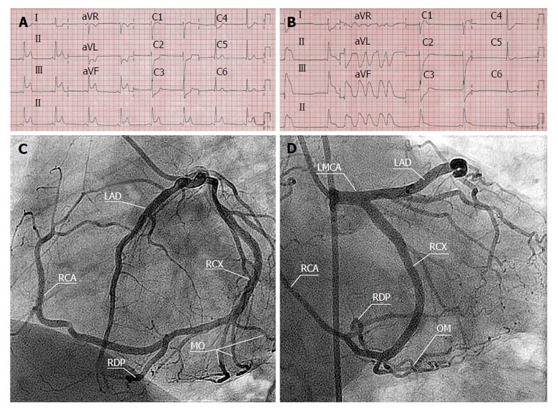
Figure 1 Resting electrocardiograph.
A: Electrocardiograph during chest pain depicting transmural ischemia in the infero-posterior leads; B: Followed by a non-sustained monomorphic ventricular tachycardia; C: Coronary angiography showed absence of the right coronary ostium and a single coronary artery arising from the left sinus of Valsalva with normal origin of the left coronary artery (LCA) having normal anatomical course of the left main stem, the left anterior descending, and the circumflex artery (Lipton L-I); D: The LCA supplies the entire myocardial tissue. No significant stenoses were found. The right coronary artery (RCA) appeared as a continuation of the distal left circumflex artery to the right atrioventricular groove and terminated near the RSV (Lipton L-I). LAD: Left anterior descending; LMCA: Left main coronary artery; RCX: Ramus circumflexus; RDP: Ramus descending posterior; OM: Obtuse marginal.
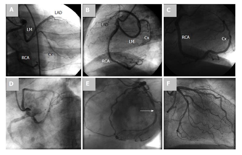
Figure 2 Coronary angiography frame.
A: Coronary angiography frame of right anterior oblique projection with cranial angulation; B: Left lateral (LL) projection showing a single origin of the right and left coronary arteries from a common right coronary ostium (Lipton R-IIP), the long curved left main stem and right dominancy are delineated; C: Coronary angiography frame in LL projection demonstrating a single coronary artery originating from the right sinus of Valsalva (RSV) giving the left anterior descending (LAD) and continued as the circumflex artery (Lipton R-I); D: Coronary angiography frame in left anterior oblique view demonstrating a single coronary artery arising from RSV as a single unique ostium (Lipton R-III); E: Coronary angiography frame in left anterior oblique view showing a single coronary artery originating from the left sinus of Valsalva. The terminal branch of circumflex artery represented the right coronary artery (Lipton L-I). Significant stenosis of the mid circumflex artery is demonstrated (white arrow); F: Coronary angiography frame demonstrates appearance of both right and left coronary arteries on injection of left sinus of Valsalva, as a single common ostium (Lipton L-IIA). Cx: Circumflex artery; RCA: Right coronary artery.
Figure 3 Angiography.
A: Supravalvular aortogram in left anterior oblique projection illustrating a single origin of the coronary arteries originating from the right sinus of Valsalva (Lipton R-IIA); B: Selective coronary angiography frame in left anterior oblique view showing a single coronary artery from the right sinus of Valsalva. Cx: Circumflex artery; LAD: Left anterior descending; RCA: Right coronary artery.
Figure 4 Transverse Multi-slice computed tomography scan in subtype (Lipton R-I) demonstrating the origin of the single coronary artery arising from the right sinus of Valsalva supplying the whole heart.
Figure 5 Volume-rendered image in subtype (Lipton L-IIB) demonstrating the inter-arterial course of the right coronary artery between the aorta and pulmonary artery.
Figure 6 Three-dimensional volume-rendered image in subtype (Lipton L-IIB) demonstrating the inter-arterial course of the right coronary artery (long arrow) between the aorta (arrowhead) and semitransparent pulmonary artery (short arrows).
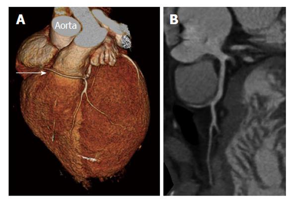
Figure 7 Single coronary artery.
A: Three-dimensional volume-rendered image of benign course of right coronary artery (arrow) from left sinus of Valsalva (Lipton L-IIA); B: Transverse multi-slice computed tomography scan in subtype (Lipton L-IIA) demonstrating the origin of the single coronary artery arising from the left sinus of Valsalva supplying the whole heart.
- Citation: Said SA, de Voogt WG, Bulut S, Han J, Polak P, Nijhuis RL, op den Akker JW, Slootweg A. Coronary artery disease in congenital single coronary artery in adults: A Dutch case series. World J Cardiol 2014; 6(4): 196-204
- URL: https://www.wjgnet.com/1949-8462/full/v6/i4/196.htm
- DOI: https://dx.doi.org/10.4330/wjc.v6.i4.196


