Copyright
©2014 Baishideng Publishing Group Co.
World J Cardiol. Apr 26, 2014; 6(4): 183-195
Published online Apr 26, 2014. doi: 10.4330/wjc.v6.i4.183
Published online Apr 26, 2014. doi: 10.4330/wjc.v6.i4.183
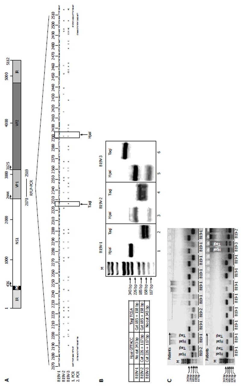
Figure 1 Generation of B19V-genotype 1 to 3 specific restriction fragment length polymorphism-polymerase chain reaction.
A: Schematic representation of the B19V genome showing localization of the B19V-genotype-specific restriction fragment length polymorphism-polymerase chain reaction (RFLP-PCR) in the B19V NS1-VP1u region. Sequences of B19V-1, B19V-2 and B19V-3 showing the RFLP-PCR fragment and HpaI and TaqI restriction enzyme sites (lower panel). Primer positions for 1st and 2nd RFLP-PCR are indicated (1. PCR and 2. PCR, see also Table 2); B: Expected fragment size and digestion pattern after HpaI and TaqI digestions (right panel). Agarose gel electrophoresis showing respective PCR-fragments after HpaI and TaqI digestion for each B19V-genotype (left panel); C: Representative agarose gel electrophoresis of patient-specific B19V RFLP-PCRs. B19V-1: B19V-genotype 1.
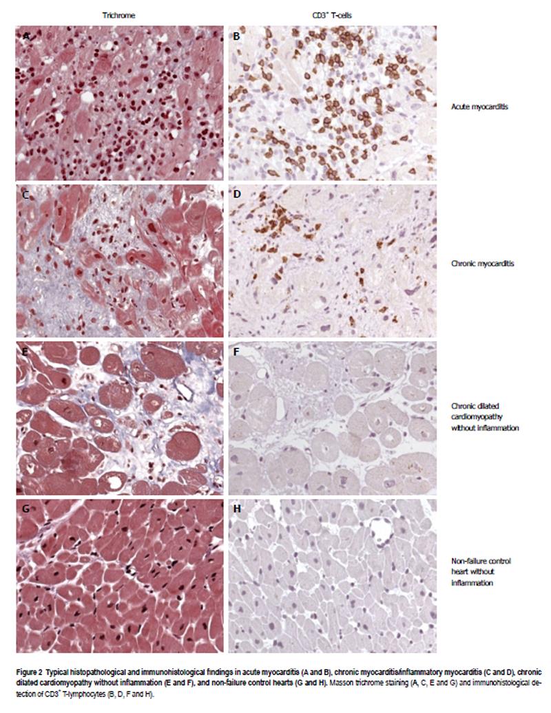
Figure 2 Typical histopathological and immunohistological findings in acute myocarditis (A and B), chronic myocarditis/inflammatory myocarditis (C and D), chronic dilated cardiomyopathy without inflammation (E and F), and non-failure control hearts (G and H).
Masson trichrome staining (A, C, E and G) and immunohistological detection of CD3+ T-lymphocytes (B, D, F and H).
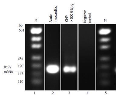
Figure 3 Representative B19V-specific reverse transcription-polymerase chain reaction showing B19V mRNA replication intermediates isolated from endomyocardial biopsies of patients with acute myocarditis (lane 2) and chronic myocarditis/iCMP (lane 3).
iCMP: Inflammatory cardiomyopathy.
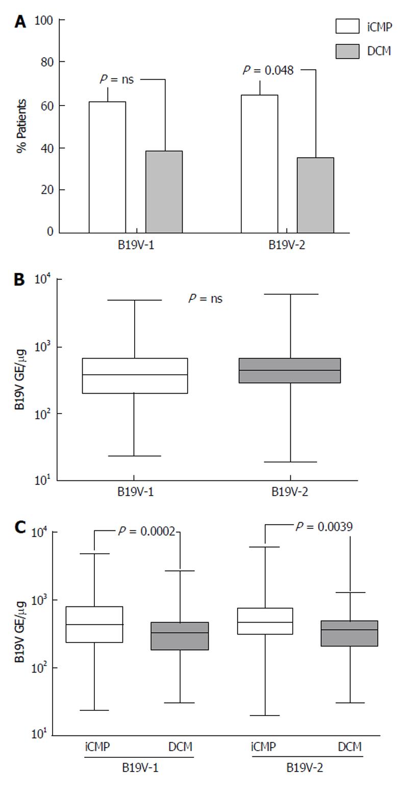
Figure 4 Genotype specific myocardial B19V loads of patients with chronic myocarditis.
A: Prevalence of B19V-genotype 1 (B19V-1) and B19V-2 in endomyocardial biopsies of patients with myocarditis [inflammatory cardiomyopathy (iCMP), grey columns] and dilated cardiomyopathy (DCM, white columns). Patient number is given in %; B: qPCR of myocardial of B19V-1 and B19V-2 loads in endomyocardial biopsies (EMBs); C: B19V genotype-specific myocardial viral loads in EMBs of patients with chronic myocarditis (iCMP, white columns) and DCM (grey columns) determined by qPCR. One-way Anova was highly significant (P < 0.0001). P < 0.05 is statistically significant (two-tailed T-test). qPCR: Quantitative real-time polymerase chain reaction.
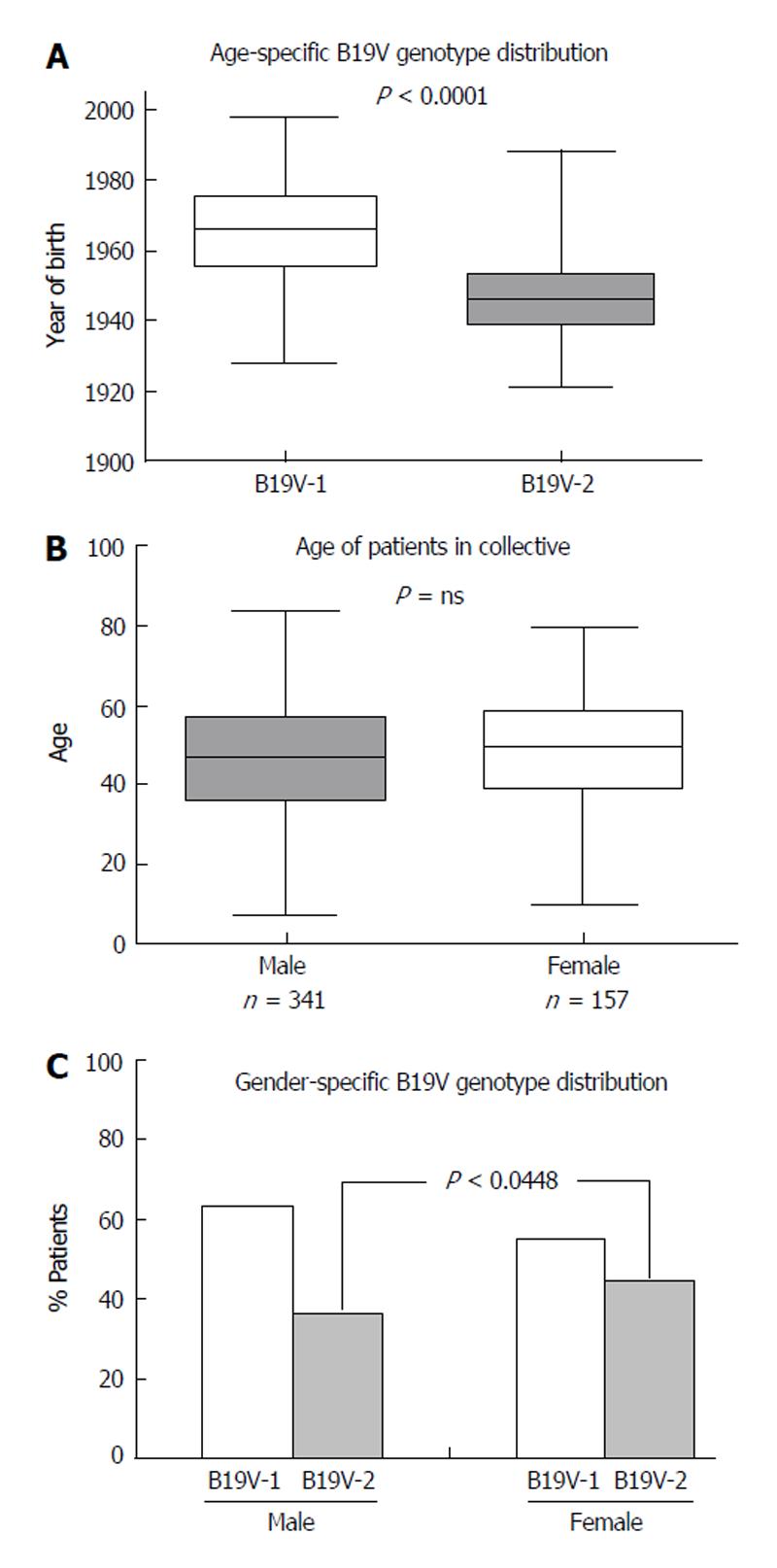
Figure 5 Age and gender dependent distribution of B19V-genotypes in endomyocardial biopsies of patients with myocarditis.
A: Distribution of B19V-genotype 1 (B19V-1) and B19V-2 according to year of birth; B: Gender-specific mean age of our patient cohort; C: Gender-specific distribution of B19V-1 (white columns) and B19V-2 (grey columns). P < 0.05 is statistically significant (two-tailed T-test).
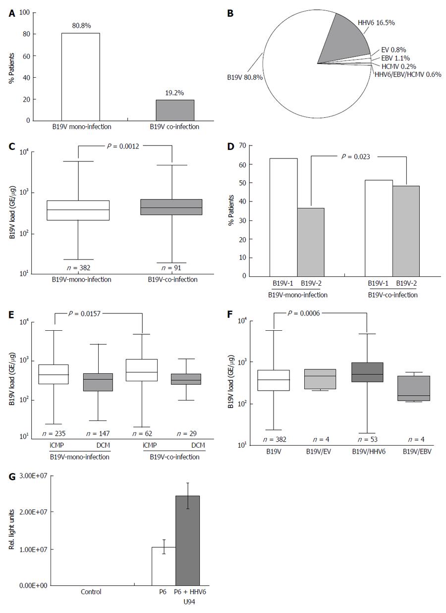
Figure 6 Distribution of B19V-coinfection with cardiotropic viruses.
A: In endomyocardial biopsies determined by virus-specific nPCR; B: Frequency of B19V-coinfection with cardiotropic viruses; C: qPCR of B19V loads in B19V mono- and co-infection; D: Distribution of B19V-genotype 1 and 2 in B19V mono- and co-infection in endomyocardial biopsies (EMBs); E: B19V loads of B19V mono- and co-infection in EMBs of patients with iCMP and DCM. One-way Anova was highly significant (P < 0.0001); F: B19V loads of B19V mono- and co-infection with cardiotropic viruses. One-way Anova was highly significant (P = 0.0091); G: Luciferase reporter assay to determine transactivation capacity of the HHV6-U94 transactivator on the B19V P6-promoter activity. P < 0.05 is statistically significant (two-tailed T-test). HHV6: Human herpesvirus 6; EV: Enterovirus; HCMV: Human cytomegalovirus; EBV: Epstein-Barr virus; DCM: Dilated cardiomyopathy; iCMP: Inflammatory cardiomyopathy; qPCR: Quantitative real-time polymerase chain reaction.
- Citation: Bock CT, Düchting A, Utta F, Brunner E, Sy BT, Klingel K, Lang F, Gawaz M, Felix SB, Kandolf R. Molecular phenotypes of human parvovirus B19 in patients with myocarditis. World J Cardiol 2014; 6(4): 183-195
- URL: https://www.wjgnet.com/1949-8462/full/v6/i4/183.htm
- DOI: https://dx.doi.org/10.4330/wjc.v6.i4.183


