Copyright
©2014 Baishideng Publishing Group Co.
World J Cardiol. Mar 26, 2014; 6(3): 112-114
Published online Mar 26, 2014. doi: 10.4330/wjc.v6.i3.112
Published online Mar 26, 2014. doi: 10.4330/wjc.v6.i3.112
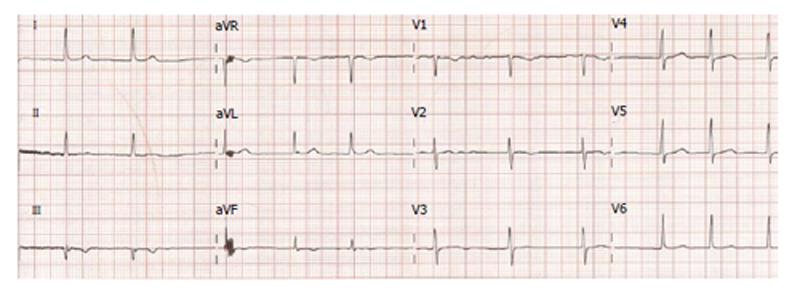
Figure 1 The patient’s electrocardiography, demonstrating new anterior T wave inversion.
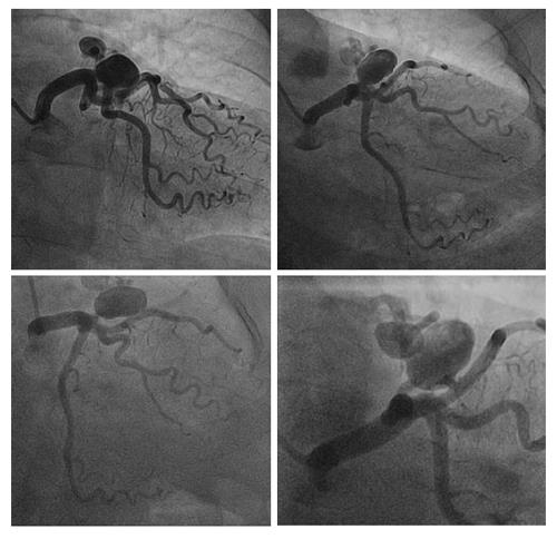
Figure 2 Coronary angiogram images demonstrating two fistulae arising from distal left main coronary artery and proximal left anterior descending artery supplying a large aneurysm.
The aneurysm drains into the pulmonary artery through the arteriovenous fistulae.
- Citation: Castles AV, Mogilevski T, Haq MAU. Steal syndrome secondary to coronary artery fistulae associated with giant aneurysm. World J Cardiol 2014; 6(3): 112-114
- URL: https://www.wjgnet.com/1949-8462/full/v6/i3/112.htm
- DOI: https://dx.doi.org/10.4330/wjc.v6.i3.112


