Copyright
©2014 Baishideng Publishing Group Co.
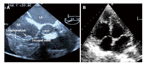
Figure 1 Transesophageal and transthoracic echocardiography before and after chemotherapy.
A: Transesophageal echocardiography showed the appearance of the lymphomatous infiltration (dotted line) and a neoplastic infiltration on the tricuspid valve (short arrow). A moderate pericardial effusion is seen; B: This transthoracic echocardiography performed after 6 cycles of rituximab, cyclophosphamide, vincristine, doxorubicin and prednisolone regime showed the resolution of the tumor mass and the disappearance of the tricuspid valve tumor infiltration; RA: Right atrium; LA: Left atrium; LVOT: Left ventricular outflow tract.
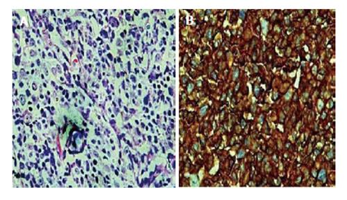
Figure 2 Histopathological staining of the left axillary lymph node.
A: The H E slide showing diffuse malignant large lymphoid cells consisting of centroblasts, immunoblasts and large centrocyte-like cells with occasional multinucleated cells (HE stain, × 40); B: The tumor cells are positive for CD20 (Immunochemistry staining, × 10).
- Citation: Abdullah HN, Nowalid WKWM. Infiltrative cardiac lymphoma with tricuspid valve involvement in a young man. World J Cardiol 2014; 6(2): 77-80
- URL: https://www.wjgnet.com/1949-8462/full/v6/i2/77.htm
- DOI: https://dx.doi.org/10.4330/wjc.v6.i2.77


