Copyright
©2013 Baishideng Publishing Group Co.
World J Cardiol. Sep 26, 2013; 5(9): 369-372
Published online Sep 26, 2013. doi: 10.4330/wjc.v5.i9.369
Published online Sep 26, 2013. doi: 10.4330/wjc.v5.i9.369
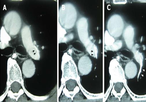
Figure 1 Contrast-enhanced computed tomography showing the proximal end of the catheter lodged in the wall of the left pulmonary artery trunk (A and B); the distal catheter end was in the small branch of left pulmonary artery (C).
The broken catheter is indicated by arrows.
Figure 2 Snare with a suture technique and the retrieved catheter.
A: A snare with a suture was curved downward by pulling the string; B: The retrieved catheter.
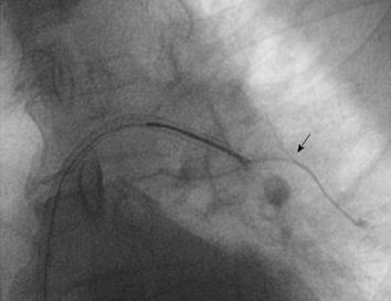
Figure 3 Image of the procedure.
The second attempt using the snare with a suture technique was successful to grasp of the body of broken catheter. The broken catheter is indicated by an arrow.
- Citation: Teragawa H, Sueda T, Fujii Y, Takemoto H, Toyota Y, Nomura S, Nakagawa K. Endovascular technique using a snare and suture for retrieving a migrated peripherally inserted central catheter in the left pulmonary artery. World J Cardiol 2013; 5(9): 369-372
- URL: https://www.wjgnet.com/1949-8462/full/v5/i9/369.htm
- DOI: https://dx.doi.org/10.4330/wjc.v5.i9.369


