Copyright
©2013 Baishideng Publishing Group Co.
World J Cardiol. Sep 26, 2013; 5(9): 364-368
Published online Sep 26, 2013. doi: 10.4330/wjc.v5.i9.364
Published online Sep 26, 2013. doi: 10.4330/wjc.v5.i9.364
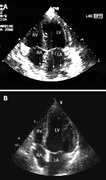
Figure 1 Transthoracic echocardiography - 4-chamber view.
A: Before chemotherapy, the echocardiogram showed tumor infiltration of the myocardium of both atria and the atrial septum (see arrow); B: After chemotherapy, the echocardiogram showed total regression of myocardial infiltration. RA: Right atrium; RV: Right ventricle; LA: Left atrium; LV: Left ventricle.
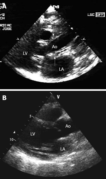
Figure 2 Transthoracic echocardiography - parasternal long-axis view.
A: Before chemotherapy, the echocardiogram showed tumor infiltration of the aorta (see arrows). B: After chemotherapy, the echocardiogram showed total regression of aortic infiltration. LA: Left atrium; LV: Left ventricle; Ao: Aorta.
- Citation: Vinicki JP, Cianciulli TF, Farace GA, Saccheri MC, Lax JA, Kazelian LR, Wachs A. Complete regression of myocardial involvement associated with lymphoma following chemotherapy. World J Cardiol 2013; 5(9): 364-368
- URL: https://www.wjgnet.com/1949-8462/full/v5/i9/364.htm
- DOI: https://dx.doi.org/10.4330/wjc.v5.i9.364


