Copyright
©2013 Baishideng Publishing Group Co.
World J Cardiol. Sep 26, 2013; 5(9): 347-354
Published online Sep 26, 2013. doi: 10.4330/wjc.v5.i9.347
Published online Sep 26, 2013. doi: 10.4330/wjc.v5.i9.347
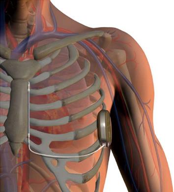
Figure 1 The subcutaneous implantable cardioverter-defibrillator system with the pulse generator implanted subcutaneously in a left lateral position, and the parasternal lead-electrode positioned parallel to and 1 to 2 cm to the left of the sternal midline.
The lead-electrode contains two sensing electrodes separated by an 8 cm shocking coil.
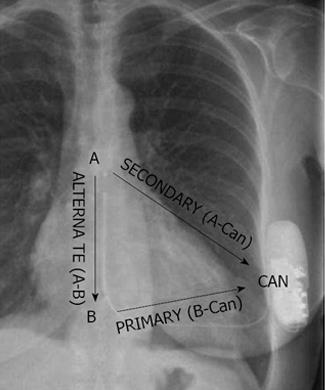
Figure 2 Chest X-ray of a patient with a subcutaneous implantable cardioverter-defibrillator system.
The cardiac rhythm is detected by 1 of the 3 available vectors, formed between the 2 sensing electrodes and the pulse generator: B-Can, A-Can, and A-B. B-Can: Proximal-to-can; A-Can: Distal-to-can; A-B: Distal to proximal.
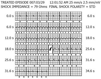
Figure 3 Subcutaneous implantable cardioverter-defibrillator electrogram showing a sustained monomorphic ventricular tachycardia that is terminated by a shock (lightning symbol).
The subcutaneous implantable cardioverter-defibrillator (S-ICD) system uses an 18/24 interval criterion for tachycardia detection (T) which is reconfirmed after capacitor charging (C), but before shock delivery, to exclude the presence of non-sustained tachyarrhythmias. S: Sensed event not classified as tachycardia.
- Citation: Akerström F, Arias MA, Pachón M, Puchol A, Jiménez-López J. Subcutaneous implantable defibrillator: State-of-the art 2013. World J Cardiol 2013; 5(9): 347-354
- URL: https://www.wjgnet.com/1949-8462/full/v5/i9/347.htm
- DOI: https://dx.doi.org/10.4330/wjc.v5.i9.347


