Copyright
©2013 Baishideng Publishing Group Co.
World J Cardiol. Sep 26, 2013; 5(9): 329-336
Published online Sep 26, 2013. doi: 10.4330/wjc.v5.i9.329
Published online Sep 26, 2013. doi: 10.4330/wjc.v5.i9.329
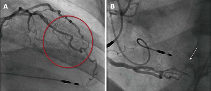
Figure 1 From the distal segment.
A: The left anterior descending coronary artery/diagonal branch multiple micro-fistulas (red circle) to the left ventricle (LV) lumen are visible; B: The right coronary artery multiple fistulas (arrow) to the LV cavity. Dual endocardial pacing leads are appreciated.
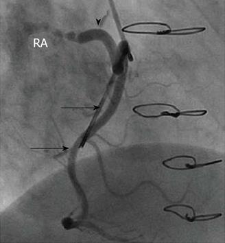
Figure 2 Dilated fistulous vessel (arrow head) originating from the proximal segment of the right coronary artery (solid arrow) and terminating into the right atrium.
The mitral valve ring is visible (hollow arrow). RA : Right atrium.
- Citation: Said SA, Schiphorst RH, Derksen R, Wagenaar L. Coronary-cameral fistulas in adults (first of two parts). World J Cardiol 2013; 5(9): 329-336
- URL: https://www.wjgnet.com/1949-8462/full/v5/i9/329.htm
- DOI: https://dx.doi.org/10.4330/wjc.v5.i9.329


