Copyright
©2013 Baishideng Publishing Group Co.
World J Cardiol. Aug 26, 2013; 5(8): 313-316
Published online Aug 26, 2013. doi: 10.4330/wjc.v5.i8.313
Published online Aug 26, 2013. doi: 10.4330/wjc.v5.i8.313
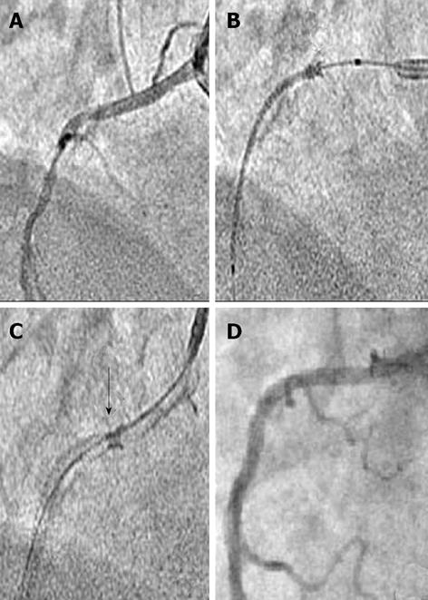
Figure 1 Percutaneous intervention of mid right coronary artery in case 1.
A: 90% eccentric, calcified, type C lesion of mid right coronary artery (RCA); B: Longitudinal compression of un-inflated stent in proximal RCA as marked by a black arrow; C: Post stent deployment, the longitudinally compressed proximal part of stent as marked by a black arrow; D: Final result showing thrombolysis in myocardial infarction-3 flow in RCA.
Figure 2 Percutaneous intervention of mid right coronary artery in case 2.
A: 90% eccentric, calcified, type C lesion of proximal right coronary artery (RCA); B: Longitudinal compression of un-inflated stent in proximal RCA as marked by a black arrow; C: Post stent deployment, the longitudinally compressed proximal part of stent as marked by a black arrow; D: Final result showing thrombolysis in myocardial infarction-3 flow in RCA.
- Citation: Vijayvergiya R, Kumar A, Shrivastava S, Kamana NK. Longitudinal stent compression of everolimus-eluting stent: A report of 2 cases. World J Cardiol 2013; 5(8): 313-316
- URL: https://www.wjgnet.com/1949-8462/full/v5/i8/313.htm
- DOI: https://dx.doi.org/10.4330/wjc.v5.i8.313


