Copyright
©2013 Baishideng Publishing Group Co.
World J Cardiol. Jul 26, 2013; 5(7): 254-257
Published online Jul 26, 2013. doi: 10.4330/wjc.v5.i7.254
Published online Jul 26, 2013. doi: 10.4330/wjc.v5.i7.254
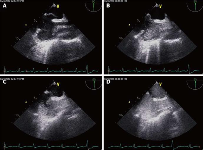
Figure 1 Transesophageal echocardiography with bubble study.
A: Transesophageal echocardiography (TEE) of the patient with a patent foramen ovale (PFO) showing a wide separation in the inter-atrial septum; B: TEE with injected agitated saline showing the presence of bubbles in the right atrium; C: TEE of the patient within the same cardiac cycle showing jet of bubbles crossing the inter-atrial septum through the PFO; D: Opacification of entire left atrium as the jet of bubbles gushes through the PFO.
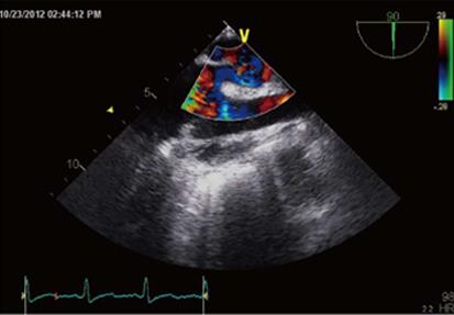
Figure 2 Transthoracic echocardiography with Doppler showing significant flow of blood across the inter-atrial septum.
- Citation: Pant S, Hayes K, Deshmukh A, Rutlen DL. Hypoxemia without persistent right-to-left pressure gradient across a patent foramen ovale: A clinical challenge. World J Cardiol 2013; 5(7): 254-257
- URL: https://www.wjgnet.com/1949-8462/full/v5/i7/254.htm
- DOI: https://dx.doi.org/10.4330/wjc.v5.i7.254


