Copyright
©2013 Baishideng Publishing Group Co.
World J Cardiol. Jun 26, 2013; 5(6): 207-209
Published online Jun 26, 2013. doi: 10.4330/wjc.v5.i6.207
Published online Jun 26, 2013. doi: 10.4330/wjc.v5.i6.207
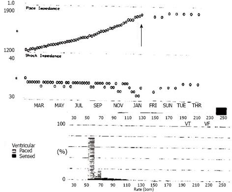
Figure 1 Automatic implantable cardioverter defibrillator interrogation showing gradually increasing pacing impedance over a period of 8 mo (arrow).
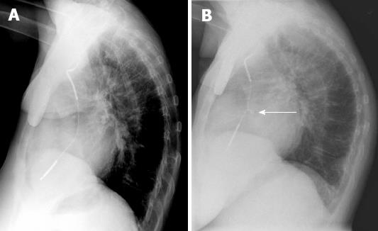
Figure 2 Chest X-ray.
A: Normal appearing lead without any twirling; B: A visible twirling is seen at right atrio-ventricular junction (arrow).
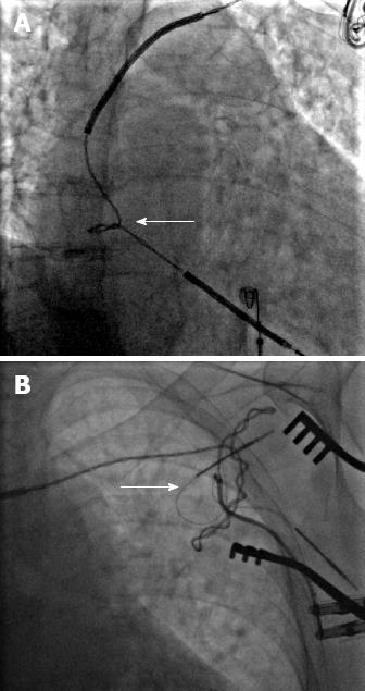
Figure 3 Antero-posterior fluoroscopic images.
A: Twisting of leads at right atrio-ventricular junction (arrow); B: Near generator in shoulder area (arrow).
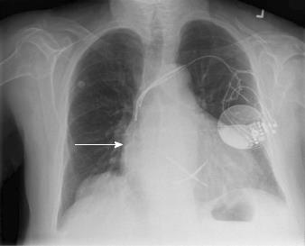
Figure 4 Antero-posterior chest X-ray after successful placement of new ventricular lead (arrow).
Abandon ventricular lead can be also be seen.
- Citation: Parikh V, Barsoum EA, Morcus R, Azab B, Lafferty J, Kohn J. Unique presentation of Twiddler’s syndrome. World J Cardiol 2013; 5(6): 207-209
- URL: https://www.wjgnet.com/1949-8462/full/v5/i6/207.htm
- DOI: https://dx.doi.org/10.4330/wjc.v5.i6.207


