Copyright
©2013 Baishideng Publishing Group Co.
World J Cardiol. Jun 26, 2013; 5(6): 175-185
Published online Jun 26, 2013. doi: 10.4330/wjc.v5.i6.175
Published online Jun 26, 2013. doi: 10.4330/wjc.v5.i6.175
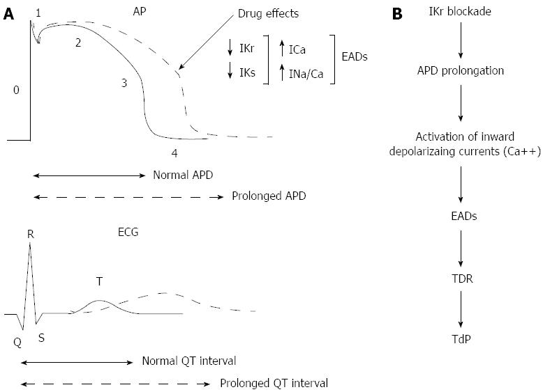
Figure 1 Relationship between the phases of ventricular transmembrane action potential and the surface electrocardiogram.
A: A reduction of outward currents (IKr, IKs) during phase 2 and 3 leads to action potential duration (APD) prolongation and QT interval prolongation. Activation of inward depolarizing currents (ICa, INa/Ca) may then give rise to early afterdepolarizations (EADs) and torsades de pointes (TdP). B: This scheme is showing that APD prolongation caused by IKr blockade is not the sole determinant for TdP. Transmural dispersion of repolarization (TDR) is required in order to form a zone of functional refractoriness in the mid myocardial layer, which is probably the basis of the re-entry that is sustaining TdP. ECG: Electrocardiographic.
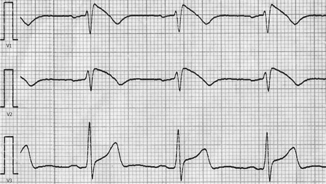
Figure 2 Diagnostic type 1 electrocardiographic pattern of Brugada syndrome.
- Citation: Konstantopoulou A, Tsikrikas S, Asvestas D, Korantzopoulos P, Letsas KP. Mechanisms of drug-induced proarrhythmia in clinical practice. World J Cardiol 2013; 5(6): 175-185
- URL: https://www.wjgnet.com/1949-8462/full/v5/i6/175.htm
- DOI: https://dx.doi.org/10.4330/wjc.v5.i6.175


