Copyright
©2013 Baishideng Publishing Group Co.
World J Cardiol. May 26, 2013; 5(5): 154-156
Published online May 26, 2013. doi: 10.4330/wjc.v5.i5.154
Published online May 26, 2013. doi: 10.4330/wjc.v5.i5.154
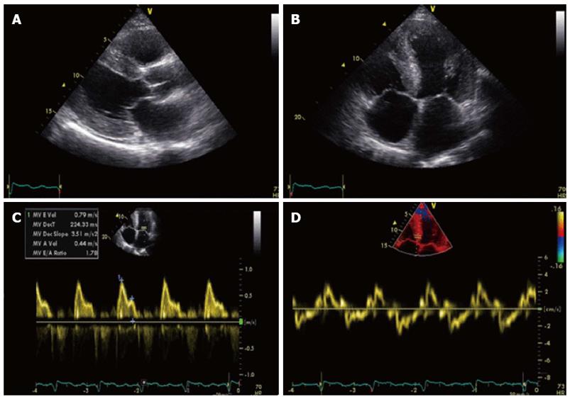
Figure 1 Transthoracic echocardiography images.
A: Parasternal long axis view diastolic still frame demonstrating thickened myocardium with sparkling of the septum. IVSd 19 mm, LPWd 19 mm; B: Apical four chamber view end diastolic still frame demonstrating thickened myocardium and normal appearance of heart valves; C: PW Doppler measurement of MV inflow. MV E/A ratio 1.8; E-vel 0.80; A-vel 0.57; IVRT 77 ms; dt 224 ms; D: Tissue Doppler Imaging with PW Doppler measurement on medial annulus of MV with E’ 3 cm/s E/E’ ratio 26.2 confirming the diastolic dysfunction. S’ 3.5 cm/s associated with impaired left ventricular function.
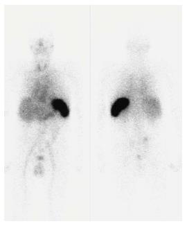
Figure 2 Serum amyloid P scintigraphy 24 h after intravenous injection of 123I-serum amyloid P.
Serum amyloid P (SAP) scintigraphy 24 h after intravenous injection of 123I-SAP. Total body uptake from the side (left image) and back (right image). Normal blood pool activity is present in organs such as liver, heart, and kidneys. Intense uptake is present in the spleen.
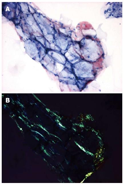
Figure 3 Abdominal subcutaneous fat aspirate of the patient stained with Congo red, magnification x 30.
Amyloid score 3+ (10%-60% of the surface is occupied by amyloid). A: When viewed in normal light, amyloid is stained red; B:The same specimen viewed in polarised light: amyloid shows apple-green birefringence.
- Citation: Brugts JJ, Houtgraaf J, Hazenberg BP, Kofflard MJ. Echocardiographic features of an atypical presentation of rapidly progressive cardiac amyloidosis. World J Cardiol 2013; 5(5): 154-156
- URL: https://www.wjgnet.com/1949-8462/full/v5/i5/154.htm
- DOI: https://dx.doi.org/10.4330/wjc.v5.i5.154


