Copyright
©2013 Baishideng Publishing Group Co.
World J Cardiol. Apr 26, 2013; 5(4): 102-105
Published online Apr 26, 2013. doi: 10.4330/wjc.v5.i4.102
Published online Apr 26, 2013. doi: 10.4330/wjc.v5.i4.102
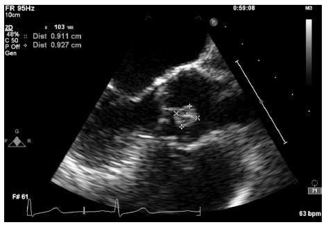
Figure 1 Mid esophageal aortic valve long axis view showing the papillary fibroelastoma attached to the aortic side of the right coronary cusp.
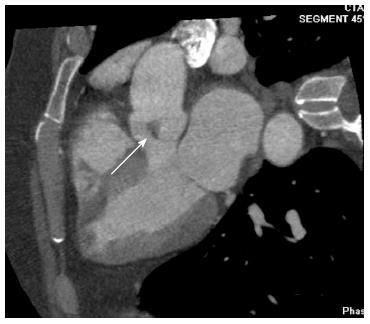
Figure 2 Cardiac computed tomography five chamber view showing the papillary fibroelastoma attached to the right coronary cusp.
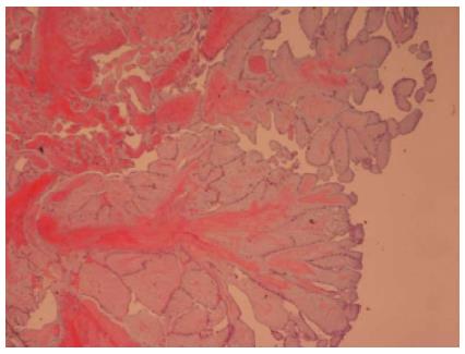
Figure 3 Histopathology.
Hematoxylin and eosin stain showing papillary fibroelastoma with narrow, elongated and branching papillary fronds with central avascular collagen and elastic tissue (Low power view, 40 × magnification).
- Citation: Aryal MR, Badal M, Mainali NR, Jalota L, Pradhan R. Papillary fibroelastoma of the aortic valve: An unusual cause of angina. World J Cardiol 2013; 5(4): 102-105
- URL: https://www.wjgnet.com/1949-8462/full/v5/i4/102.htm
- DOI: https://dx.doi.org/10.4330/wjc.v5.i4.102


