Copyright
©2013 Baishideng Publishing Group Co.
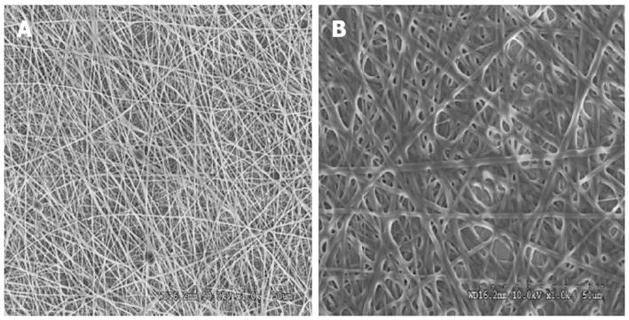
Figure 1 Scanning electron microscope image showing the fiber morphology of (A) collagen fibers with a fiber diameter of 380 ± 77 nm (B) poly (glycerol sebacate)/collagen core/shell fibers with a fiber diameter of 1192 ± 277 nm at 1000 × magnification.
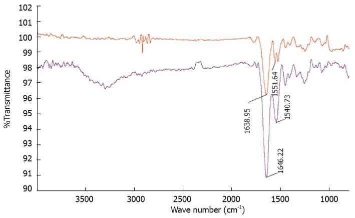
Figure 2 Fourier transform infrared spectroscopy images of red curve-collagen fibers showing characteristic amide peaks at 1638.
95 cm-1and 1551.64 cm-1 blue curve-poly (glycerol sebacate)/collagen core/shell fibers showing characteristic amide peaks at 1646.22 cm-1 and 1540.73 cm-1.
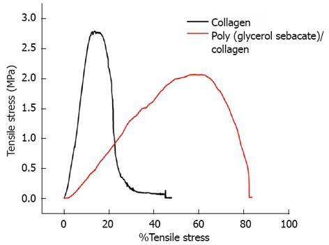
Figure 3 Tensile stress-strain curves of collagen fibers and poly (glycerol sebacate)/collagen core/shell fibers.
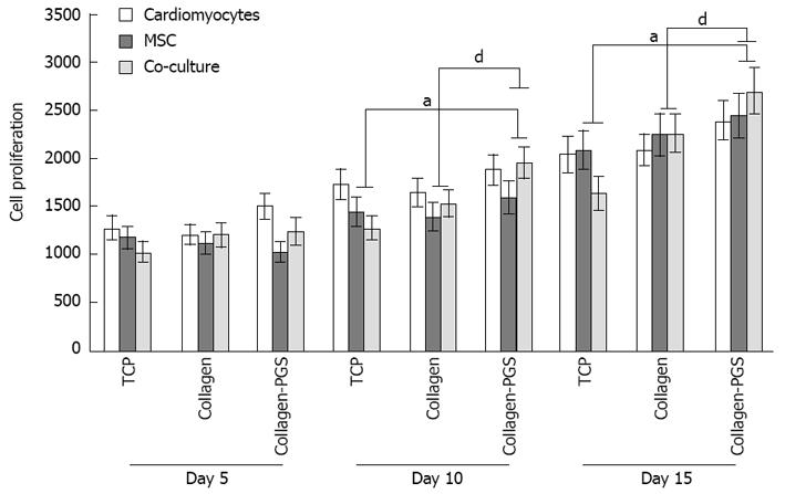
Figure 4 Cell proliferation study for days 5, 10 and 15 on tissue culture plate, poly (glycerol sebacate)/collagen core/shell fibers and collagen nanofibers using cardiomyocytes, mesenchymal stem cells and cardiomyocytes-mesenchymal stem cells co-culture.
Denotes statistical significant difference MSC aP≤ 0.05 vs co-culture cells cultured on TCP and PGS/collagen core/shell fibers; Denotes statistical significant difference MSC dP≤ 0.01 vs co-culture cells cultured on collagen and PGS/collagen core/shell fibers. TCP: Tissue culture plate; PGS: Poly (glycerol sebacate); MSCs: Mesenchymal stem cells.
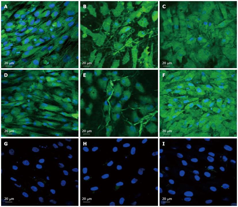
Figure 5 Immunocytochemical analysis for the expression of cardiac marker protein actinin at 60 × magnification on the tissue culture plate (A, D, G), collagen nanofibers (B, E, H) and poly (glycerol sebacate)/collagen core/shell fibers (C, F, I) comprising of cardiomyocytes (A-C), mesenchymal stem cells-cardiomyocytes co-culture group (D-F) and mesenchymal stem cells (G-I).
Nucleus stained with 4,6-diamidino-2-phenylindole hydrochloride.
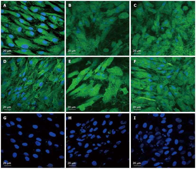
Figure 6 Immunocytochemical analysis for the expression of the cardiac marker protein Troponin at 60 × magnification on the tissue culture plate (A, D, G), collagen nanofibers (B, E, H) and poly (glycerol sebacate)/collagen core/shell fibers (C, F, I) comprising of cardiomyocytes (A-C), mesenchymal stem cells-cardiomyocytes co-culture group (D-F) and mesenchymal stem cells (G-I).
Nucleus stained with 4,6-diamidino-2-phenylindole hydrochloride.
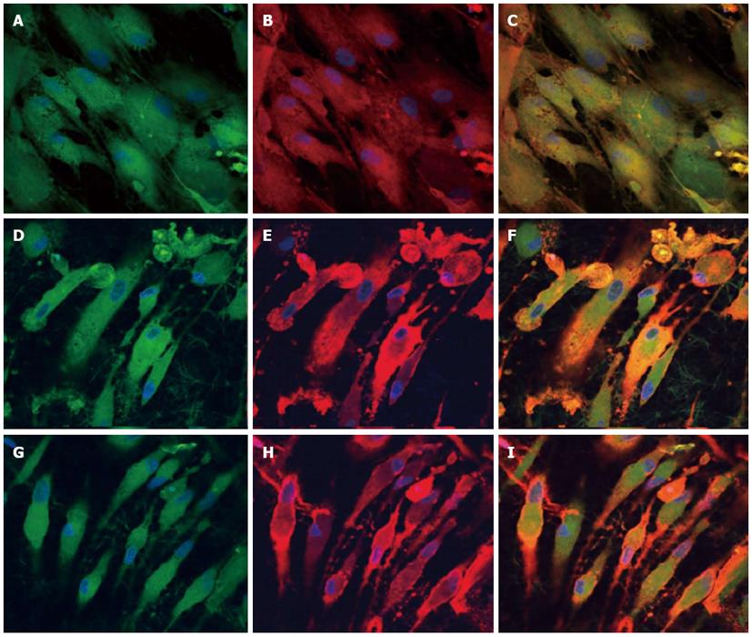
Figure 7 Dual immunocytochemical analysis for the expression of mesenchymal stem cells marker protein CD 105 (A, D, G) and cardiac marker protein Actinin (B, E, H) in the co-culture samples and the merged image showing the dual expression of both CD 105 and Actinin (C, F, I); on the tissue culture plate (A, B, C), collagen nanofibers (D, E, F) and poly (glycerol sebacate)/collagen core/shell fibers (G, H, I) at 60 × magnification.
Nucleus stained with 4,6-diamidino-2-phenylindole hydrochloride.
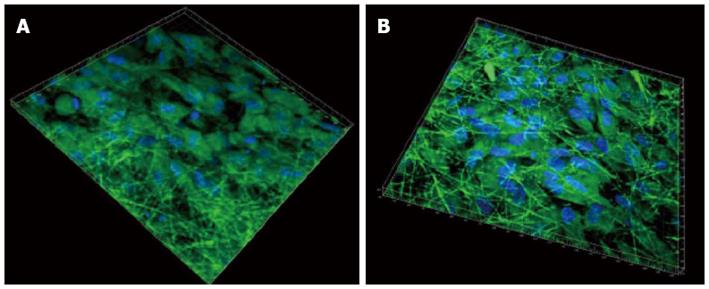
Figure 8 3D image using Imaris software of cardiomyocytes-mesenchymal stem cells co-culture group stained with cardiac specific marker protein troponin at 60 × magnification on (A) collagen fibers (B) poly (glycerol sebacate)/collagen core/shell fibers.
Nucleus stained with 4,6-diamidino-2-phenylindole hydrochloride.
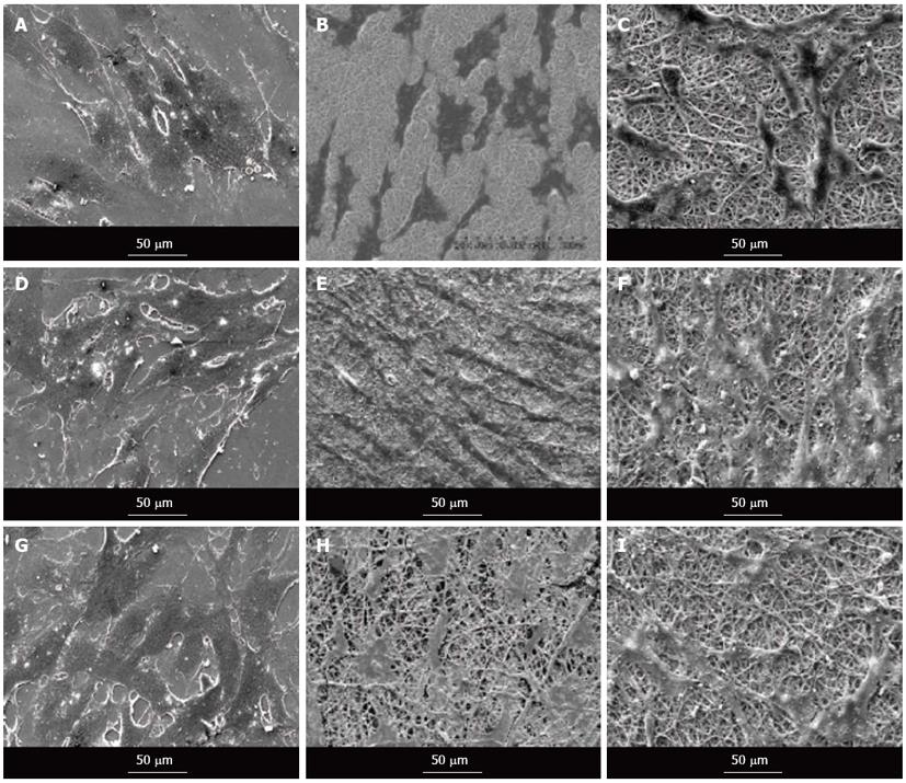
Figure 9 Scanning electron microscope images showing the cell morphology of cardiomyocytes (A-C), cardiomyocytes-mesenchymal stem cells co-culture cells (D-F) and mesenchymal stem cells (G-I) grown on tissue culture plate (A, D, G), collagen nanofibers (B, E, H) and poly (glycerol sebacate)/collagen core/shell fibers (C, F, I) on day 15 at 500 × magnification.
- Citation: Ravichandran R, Venugopal JR, Sundarrajan S, Mukherjee S, Ramakrishna S. Cardiogenic differentiation of mesenchymal stem cells on elastomeric poly (glycerol sebacate)/collagen core/shell fibers. World J Cardiol 2013; 5(3): 28-41
- URL: https://www.wjgnet.com/1949-8462/full/v5/i3/28.htm
- DOI: https://dx.doi.org/10.4330/wjc.v5.i3.28


