Copyright
©2013 Baishideng Publishing Group Co.
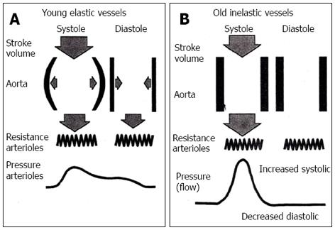
Figure 1 In a young person the elastic aorta expands during systole and absorbs part of the stroke volume.
A: This figure depicts the function of the central aorta of a younger person during systole and diastole. During systole, the elastic aorta with each cardiac stroke volume (top filled arrow) is dilated and functions as a reservoir. As a result, not all stroke volume (SV) is transmitted distally. During diastole the elastic recoil of the aorta expels the remnant original SV to distal arteries and arterioles. This function results in a smooth contour of the arterial pulse wave and a narrow pulse pressure (PP) (bottom); B: In an older person, the aorta has lost most of its elasticity resulting in a reduction of its reservoir or capacitance function, resulting in the expulsion of almost the entire SV to the distal arteries with practically no diastolic blood flow (top filled arrow. This results in a distortion of the arterial pulse wave (bottom), an increase in systolic blood pressure, a decrease in diastolic blood pressure, and a widening of PP. Adapted with permission from Franklin et al[12].
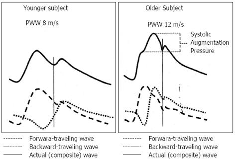
Figure 2 This figure depicts the configuration of the arterial waveforms in the younger person (left) and the older person (right).
The arterial waveforms are composite waves (top heavy line), composed of a forward traveling wave (dashed line) and a backward traveling reflective wave (dotted line). The vertical line represents the closure of the aortic valve. The top solid line indicates the peak systolic blood pressure (SBP) in the younger person (left) and the older person (right) together with the augmentation pressure. The reflected wave in the younger person (left), returns to the aortic root early in diastole augmenting the diastolic blood pressure and improving the coronary circulation. In the older person (right), the reflected wave returns to the aortic root late in systole, thus augmenting the SBP and increasing the left ventricular outflow pressure leading to left ventricular hypertrophy. Due to the arterial stiffness in the older person, the pulse wave velocity is increased (12 m/s) compared to the younger person (8 m/s). Adapted with permission from Franklin et al[12].
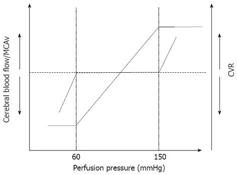
Figure 3 This figure depicts the cerebral blood flow autoregulation and the range of perfusion pressure.
An autoregulatory plateau is seen between 60 to 150 mmHg of mean arterial pressure (MAP). This autoregulatory plateau is maintained through changes in cerebral vascular resistance (CVR). Once the limits of autoregulation are reached, CVR cannot correct for further changes in pressure as demonstrated by the MAP limits of < 60 mmHg (lower limit) and > 150 mmHg (upper limit). Adapted with permission from Lucas et al[14].
- Citation: Chrysant SG. Treating blood pressure to prevent strokes: The age factor. World J Cardiol 2013; 5(3): 22-27
- URL: https://www.wjgnet.com/1949-8462/full/v5/i3/22.htm
- DOI: https://dx.doi.org/10.4330/wjc.v5.i3.22


