Copyright
©2013 Baishideng.
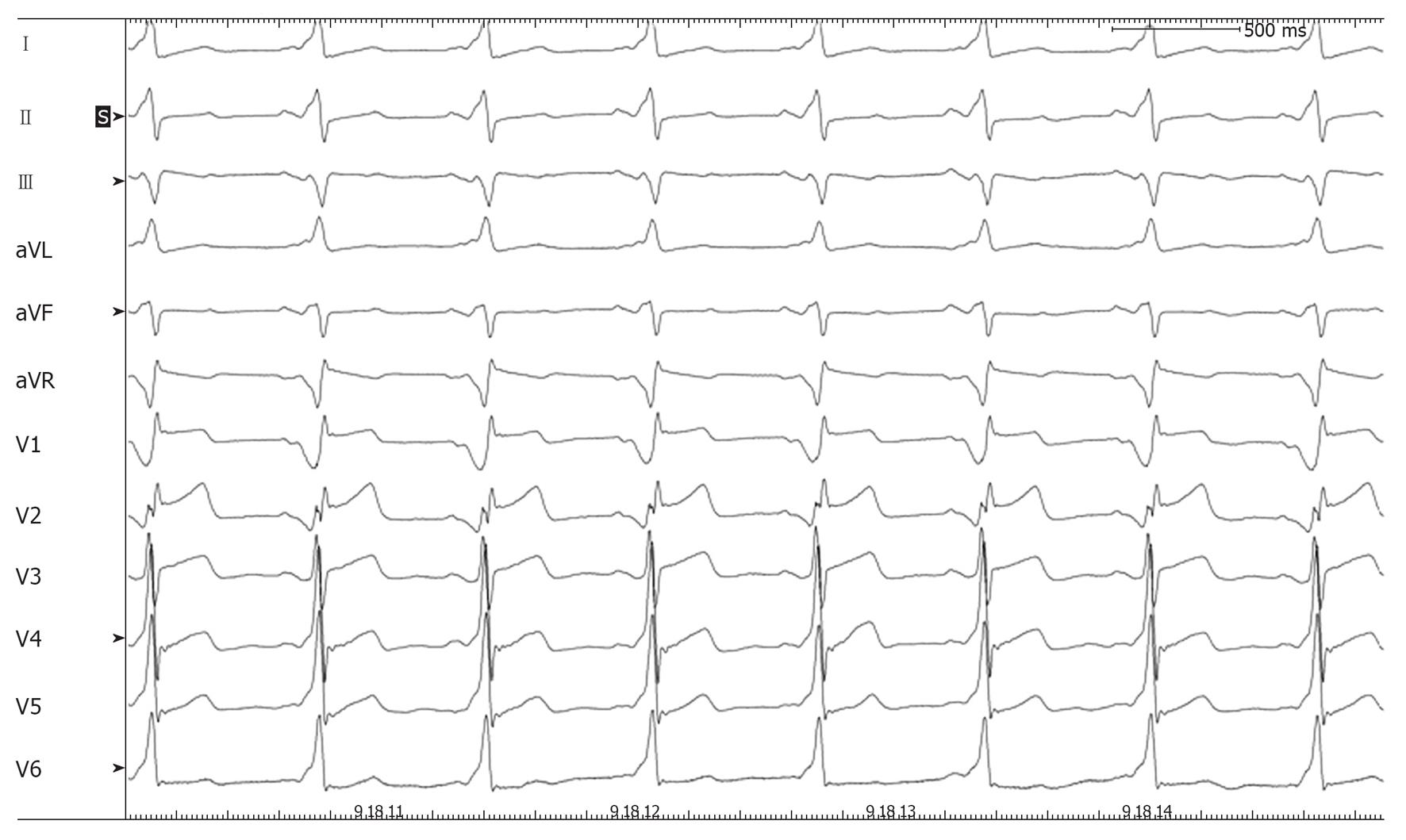
Figure 1 Basal 12-lead electrocardiograms showing constant ventricular preexcitation.
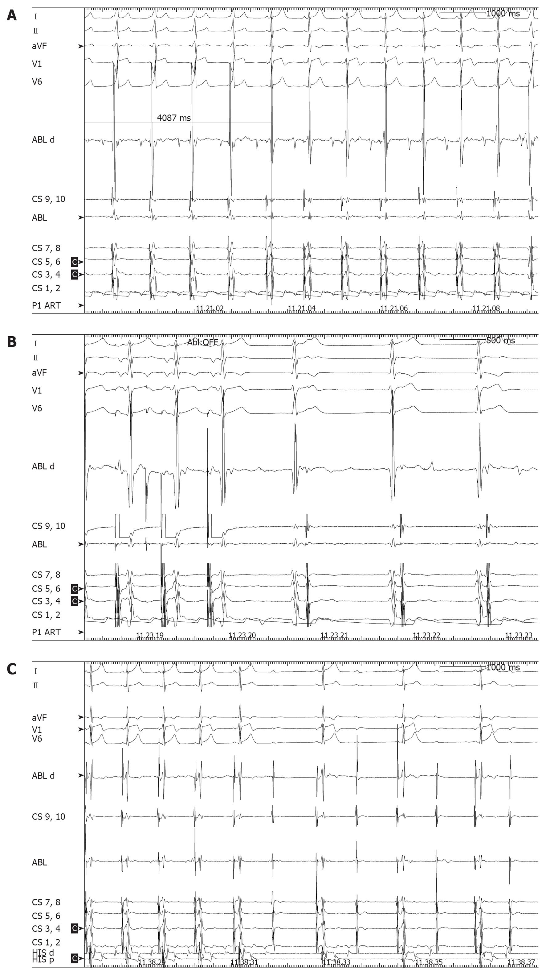
Figure 2 Three consolidation pulses were delivered at the same site.
A: The effective radiofrequency (RF) pulse. Ventricular preexcitation disappeared 4 s after the pulse was started and, few beats later, a junctional rhythm with 1:1 retrograde conduction overtook sinus rhythm. Thus the RF pulse was prematurely stopped. As the phenomenon could be reliably reproduced, subsequent consolidation pulses were delivered during atrial pacing with the irrigated-tip ablation catheter up to a maximum of 15 W; B: A RF pulse delivered during atrial pacing with emergence of junctional rhythm as pacing was stopped; C: Transient complete atrioventricular block during adenosine injection at the end of the first ablation procedure.
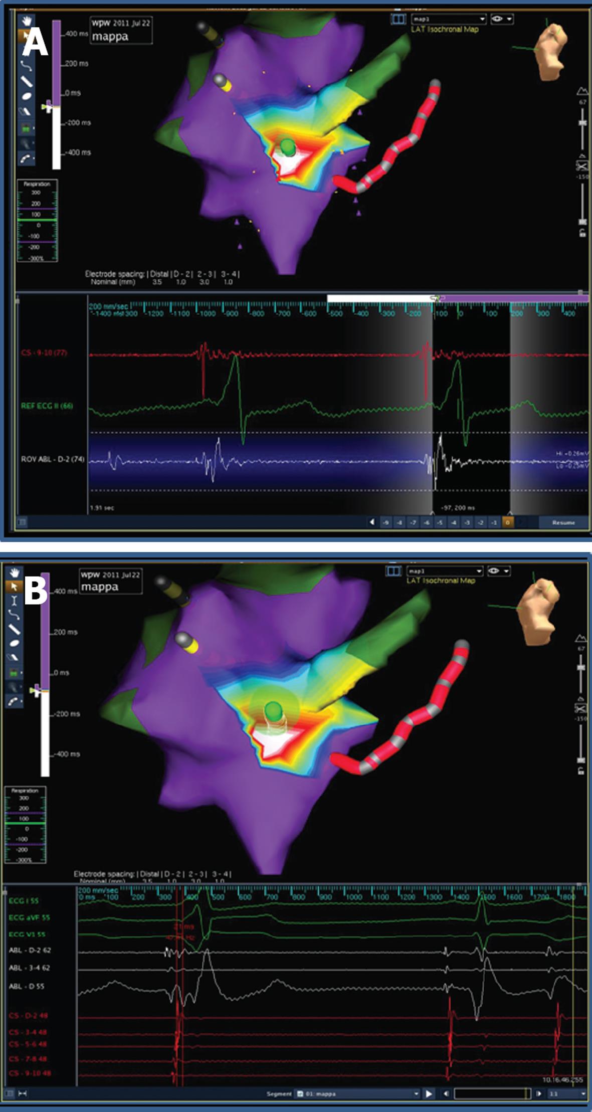
Figure 3 Two different frames obtained from the off-line analysis of geometry reconstruction recording.
A: The ablation catheter (visualized in green) is at the site of earliest ventricular activation; B: The ablation catheter is in a site slightly superior to that where mechanical “bumping” occurred.
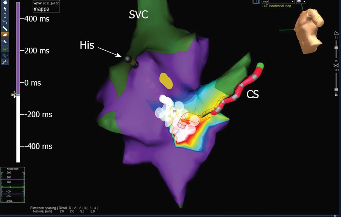
Figure 4 Site of effective ablation.
Ablation pulses (white circles) were delivered in the posterior and postero-midseptal region covering all the area where the earliest activation had been recorded and the mechanical bumping occurred. The yellow circle points out the area where the mapping catheter produced mechanical junctional beats; this area is marked as the likely site of compact atrioventricular node. Thus ablation was safely delivered up to 30 W with an irrigated tip catheter. SVC: Superior vena cava; CS: Coronary sinus.
- Citation: Casella M, Dello Russo A, Fassini G, Andreini D, De Iuliis P, Mushtaq S, Bartoletti S, Riva S, Tondo C. Manifold benefits of choosing a minimally fluoroscopic catheter ablation approach. World J Cardiol 2013; 5(2): 8-11
- URL: https://www.wjgnet.com/1949-8462/full/v5/i2/8.htm
- DOI: https://dx.doi.org/10.4330/wjc.v5.i2.8


