Copyright
©2013 Baishideng.
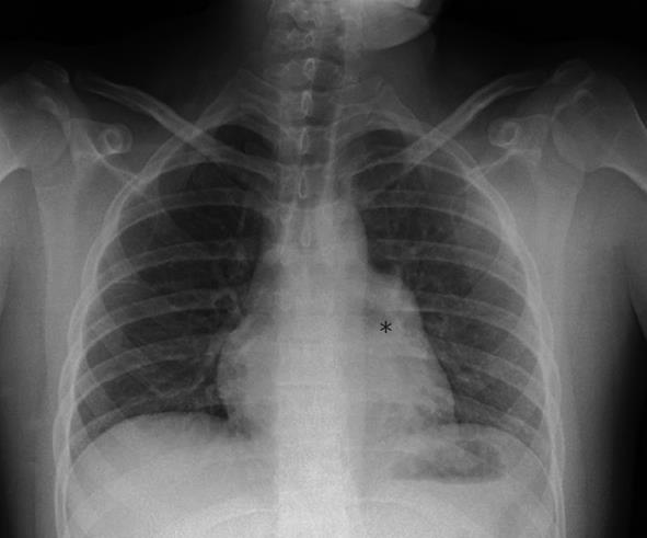
Figure 1 Chest radiograph.
Chest radiograph showing horizontalization of the left bronchus (asterisk) initially interpreted as hilar adenopathies and later found to be secondary to herniation of the left atrial appendage through the pericardial defect.
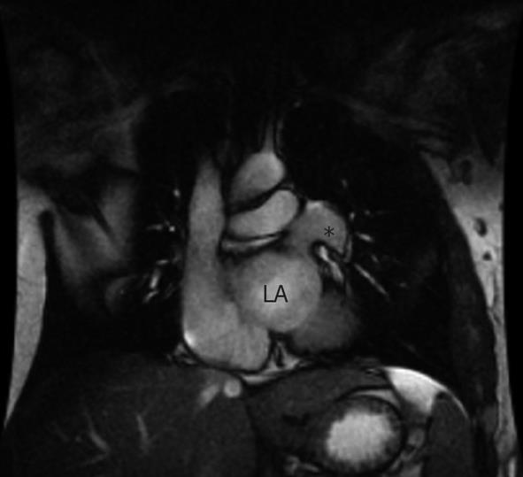
Figure 2 Cardiac magnetic resonance imaging.
Cardiac magnetic resonance imaging (coronal view) displaying the partial pericardial defect (20 mm × 30 mm) localized to the left atrial (LA) wall. Herniation of the left atrial appendage can been seen (asterisk).
- Citation: Juárez AL, Akerström F, Alguacil AM, González BS. Congenital partial absence of the pericardium in a young man with atypical chest pain. World J Cardiol 2013; 5(2): 12-14
- URL: https://www.wjgnet.com/1949-8462/full/v5/i2/12.htm
- DOI: https://dx.doi.org/10.4330/wjc.v5.i2.12


