Copyright
©2013 Baishideng Publishing Group Co.
World J Cardiol. Oct 26, 2013; 5(10): 391-393
Published online Oct 26, 2013. doi: 10.4330/wjc.v5.i10.391
Published online Oct 26, 2013. doi: 10.4330/wjc.v5.i10.391
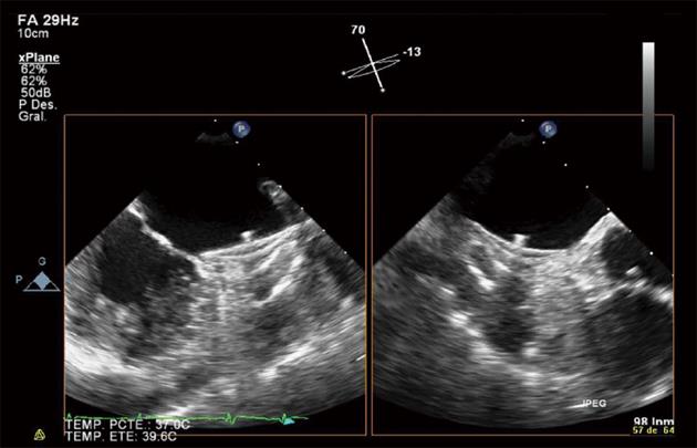
Figure 1 Forty-five day control.
Transesophageal echocardiography two-dimensional X-plane processing, simultaneous visualization at 70° (left) and 13° (right). Successful Amplatzer™ cardiac plug implantation: completely covering the left atrial appendage ostium by the occluder disk and no evidence of device thrombosis.
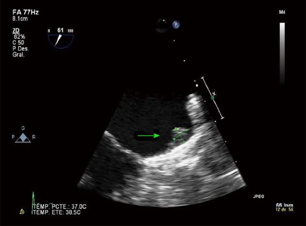
Figure 2 Four month control.
Transesophageal echocardiography two-dimensional image. Adequate covering of the left atrial appendage ostium but little thrombus (7 mm × 7 mm) is observed at the top of the button of the Amplatzer™ cardiac plug.
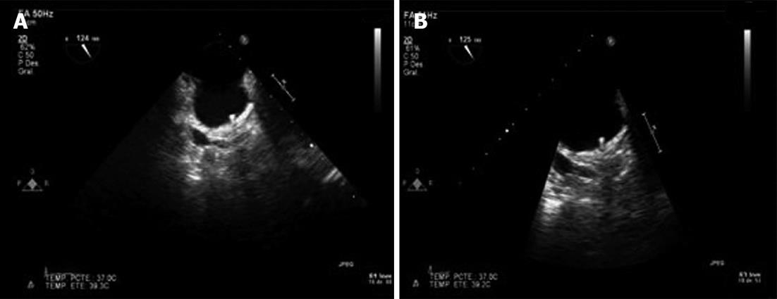
Figure 3 Transesophageal echocardiography two-dimensional image.
A: Control after 2 wk of intravenous sodium heparin treatment. Complete resolution of button thrombosis and correct device positioning; B: 6 mo control. Correct device positioning and absence of button thrombosis.
- Citation: Fernández-Rodríguez D, Vannini L, Martín-Yuste V, Brugaletta S, Robles R, Regueiro A, Masotti M, Sabaté M. Medical management of connector pin thrombosis with the Amplatzer cardiac plug left atrial closure device. World J Cardiol 2013; 5(10): 391-393
- URL: https://www.wjgnet.com/1949-8462/full/v5/i10/391.htm
- DOI: https://dx.doi.org/10.4330/wjc.v5.i10.391


