Copyright
©2012 Baishideng Publishing Group Co.
World J Cardiol. Aug 26, 2012; 4(8): 256-259
Published online Aug 26, 2012. doi: 10.4330/wjc.v4.i8.256
Published online Aug 26, 2012. doi: 10.4330/wjc.v4.i8.256
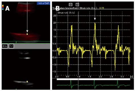
Figure 1 Measurement of the strain rate of the ascending aorta by tissue Doppler echocardiography.
A: A region of interest (arrow) was identified on the ascending aortic wall; B: Strain rate (arrow) was recorded and the peak value during initial contraction was measured by averaging the systolic peak of three heart beats.
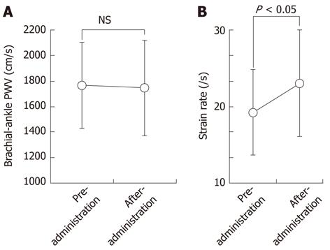
Figure 2 Effect of eicosapentaenoic acid on brachial-ankle pulse wave velocity and the strain rate of the ascending aorta.
A: No difference was noted in baPWV before and after eicosapentaenoic acid (EPA) administration; B: In contrast, a statistically significant increase in the strain rate of the ascending aorta was observed after one year of EPA administration. PWV: Pulse wave velocity; NS: Not significant.
- Citation: Haiden M, Miyasaka Y, Kimura Y, Tsujimoto S, Maeba H, Suwa Y, Iwasaka T, Shiojima I. Effect of eicosapentaenoic acid on regional arterial stiffness: Assessment by tissue Doppler imaging. World J Cardiol 2012; 4(8): 256-259
- URL: https://www.wjgnet.com/1949-8462/full/v4/i8/256.htm
- DOI: https://dx.doi.org/10.4330/wjc.v4.i8.256


