Copyright
©2012 Baishideng Publishing Group Co.
World J Cardiol. Jul 26, 2012; 4(7): 240-241
Published online Jul 26, 2012. doi: 10.4330/wjc.v4.i7.240
Published online Jul 26, 2012. doi: 10.4330/wjc.v4.i7.240
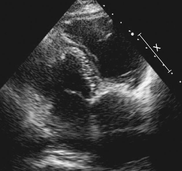
Figure 1 Echo four-chamber image of the tricuspid valve with metallic artifact making visualization of the components of the prosthesis suboptimal.
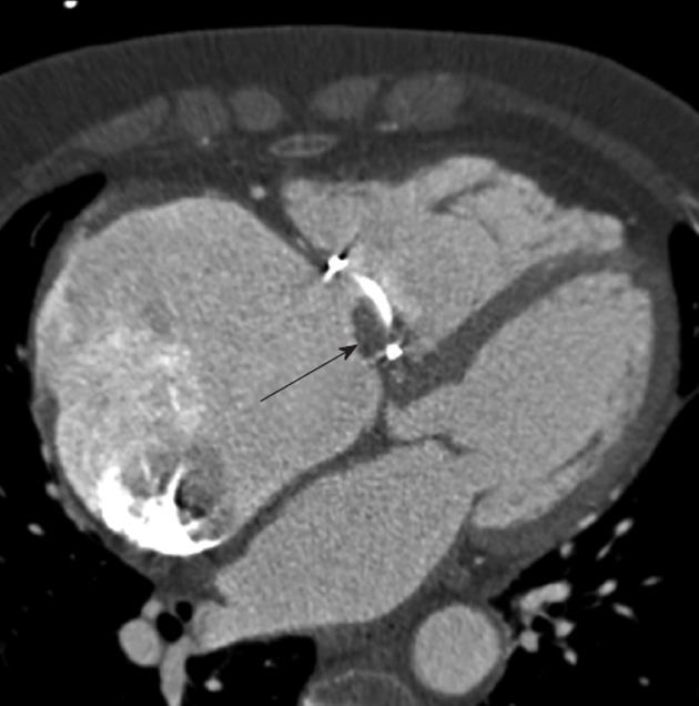
Figure 2 Coronary computed tomography image demonstrated atrialization of the right ventricle, a markedly dilated right atrium and prosthetic tricuspid valve.
Thrombus was identified between the septal flap and the valve ring of the prosthesis (arrow).
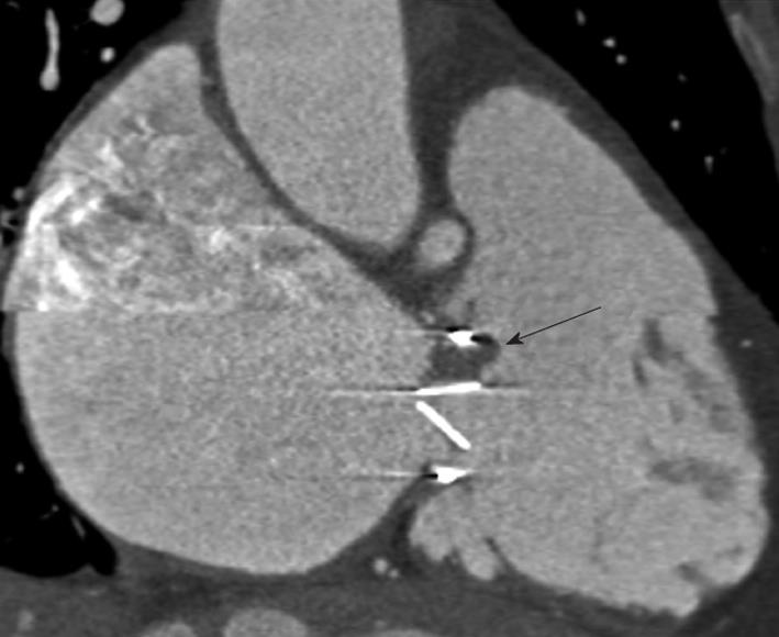
Figure 3 Cardiac computed tomography sagittal oblique multiplanar reformat demonstrated thrombus on the septal side of the prosthetic valve ring (arrow).
The right atrium was markedly dilated.
- Citation: O’Neill AC, Kelly RM, McCarthy CJ, Martos R, McCreery C, Dodd JD. Thrombosed prosthetic valve in Ebstein's anomaly: Evaluation with echocardiography and 64-slice cardiac computed tomography. World J Cardiol 2012; 4(7): 240-241
- URL: https://www.wjgnet.com/1949-8462/full/v4/i7/240.htm
- DOI: https://dx.doi.org/10.4330/wjc.v4.i7.240


