Copyright
©2012 Baishideng Publishing Group Co.
World J Cardiol. Jul 26, 2012; 4(7): 234-239
Published online Jul 26, 2012. doi: 10.4330/wjc.v4.i7.234
Published online Jul 26, 2012. doi: 10.4330/wjc.v4.i7.234
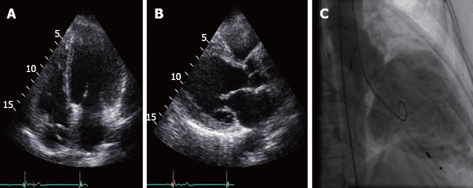
Figure 1 Transthoracic echocardiogram: apical 4-chamber view (A), parasternal long-axis (B); and (C) left ventricle angiogram showing a dilated and aneurysmatic left ventricle (36.
37 mm/m2 end diastolic and 31.44 mm/m2 end systolic diameter) with generalized hypokinesia.
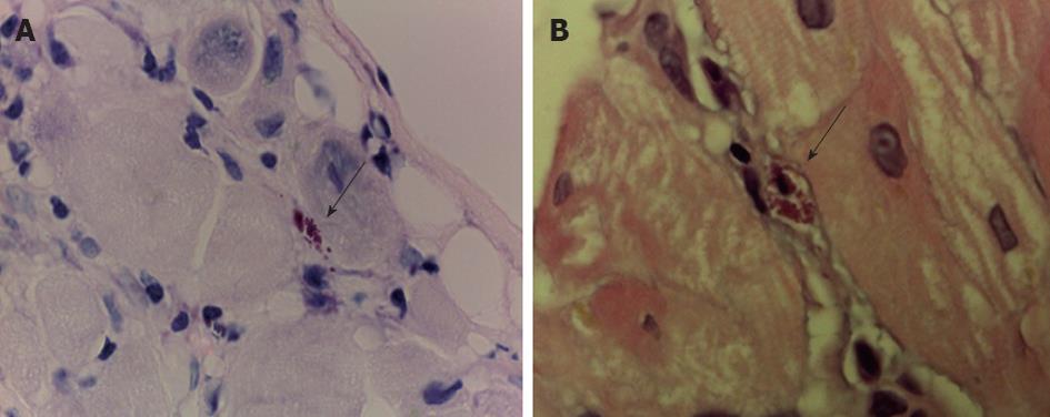
Figure 2 T.
cruzi pseudocyst (arrow). A: Heart muscle, Giemsa staining, 100×; B: Heart muscle, hematoxylin-eosin staining, 100×.
- Citation: Cortez J, Providência R, Ramos E, Valente C, Seixas J, Meruje M, Leitão-Marques A, Vieira A. Emerging and under-recognized Chagas cardiomyopathy in non-endemic countries. World J Cardiol 2012; 4(7): 234-239
- URL: https://www.wjgnet.com/1949-8462/full/v4/i7/234.htm
- DOI: https://dx.doi.org/10.4330/wjc.v4.i7.234


