Copyright
©2012 Baishideng Publishing Group Co.
World J Cardiol. Jun 26, 2012; 4(6): 214-217
Published online Jun 26, 2012. doi: 10.4330/wjc.v4.i6.214
Published online Jun 26, 2012. doi: 10.4330/wjc.v4.i6.214
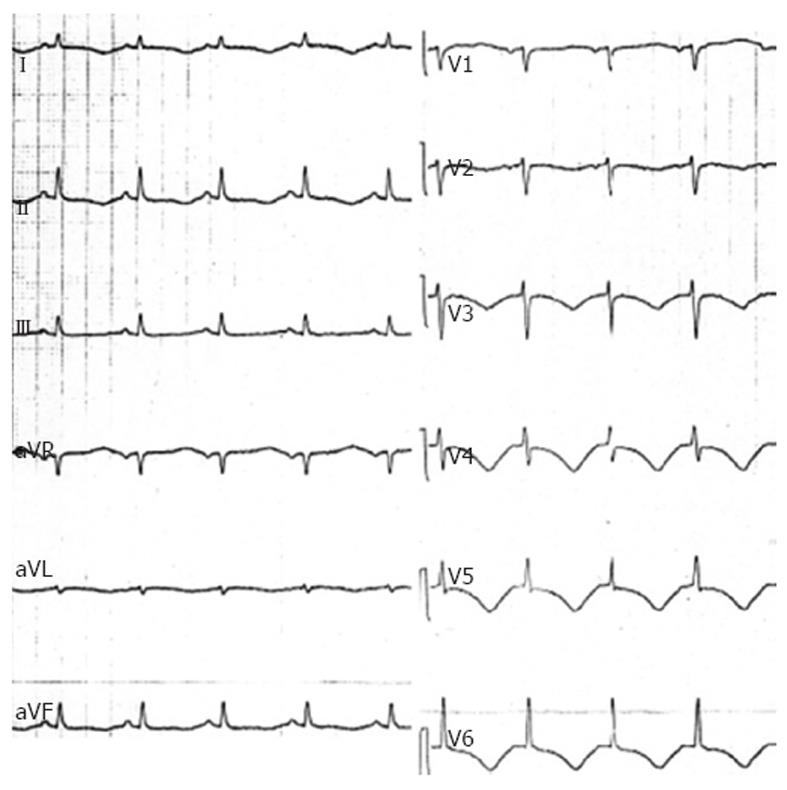
Figure 1 Electrocardiography performed in the acute setting showing diffuse T waves inversion and QT-prolongation.
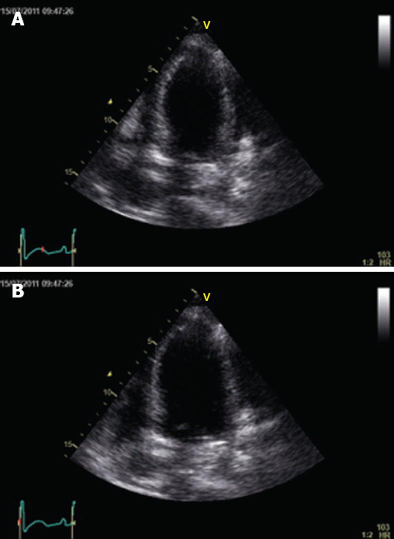
Figure 2 Baseline echocardiography recorded at the end of systole (A) and diastole (B) showing impairment of contractility of all distal-apical segments, but sparing the base and midventricular regions.
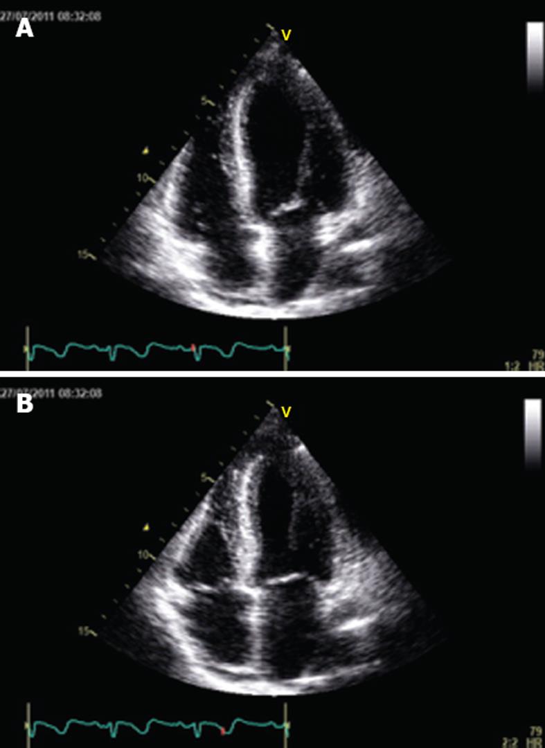
Figure 3 Echocardiography performed 3 wk later showed complete normalization of left ventricular function and resolution of contractility abnormalities, at the end of diastole (A) and at the end of systole (B).
- Citation: Bortnik M, Verdoia M, Schaffer A, Occhetta E, Marino P. Ventricular fibrillation as primary presentation of takotsubo cardiomyopathy after complicated cesarean delivery. World J Cardiol 2012; 4(6): 214-217
- URL: https://www.wjgnet.com/1949-8462/full/v4/i6/214.htm
- DOI: https://dx.doi.org/10.4330/wjc.v4.i6.214


