Copyright
©2012 Baishideng Publishing Group Co.
World J Cardiol. May 26, 2012; 4(5): 188-194
Published online May 26, 2012. doi: 10.4330/wjc.v4.i5.188
Published online May 26, 2012. doi: 10.4330/wjc.v4.i5.188
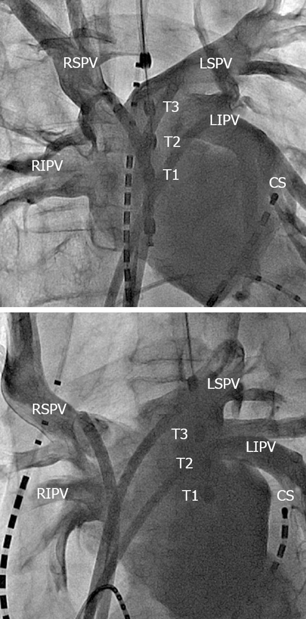
Figure 1 Left arteriography.
Fluoroscopy of left arteriography using the trans-septal sheath after short iatrogenic complete AV-block using high-frequency right ventricular stimulation. The relationship is shown of the eso-temperature probe (Eso) with three thermistor electrodes (T1-T3) to the ostium of the pulmonary veins. Upper: Right anterior oblique; Lower: Left anterior oblique. LSPV: Left superior pulmonary vein; LIPV: Left inferior pulmonary vein; RSPV: Right superior pulmonary vein; RIPV: Right inferior pulmonary vein.
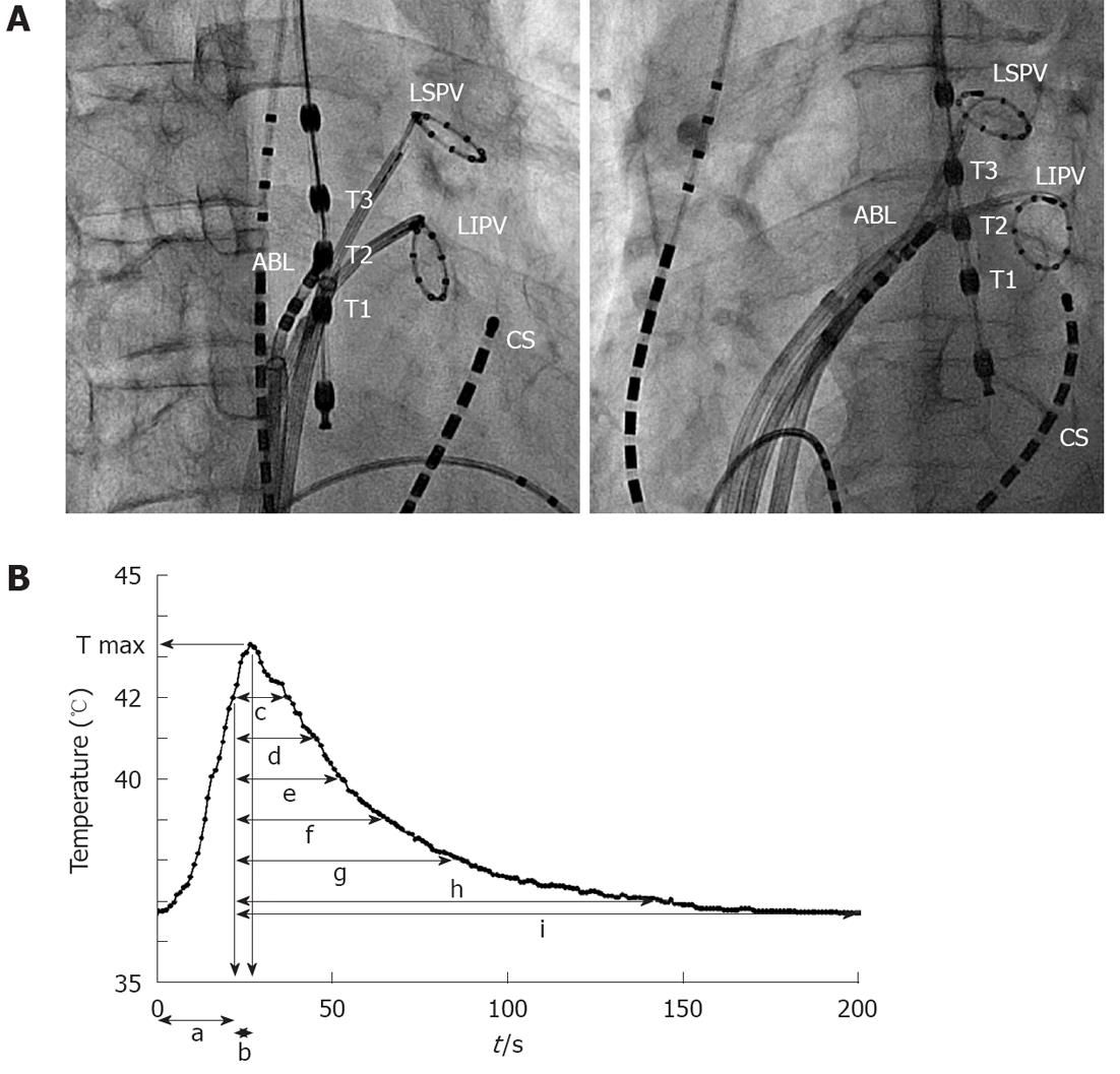
Figure 2 Position of ablation catheter (A) (left: right anterior oblique, right: left anterior oblique) and luminal esophageal temperature monitoring graph (B).
We measured the time when luminal esophageal temperature (LET) reached the cut-off temperature (a), the maximum temperature (T max) of LET, the time to reach T max after the LET reached 42 °C (b) and the time to come back from the delivery of energy end to 42 °C (c), 41 °C (d), 40 °C (e), 39 °C (f), 38 °C (g) and 37 °C (h), and the temperature before the delivery of energy (i). LSPV: Left superior pulmonary vein; LIPV: Left inferior pulmonary vein; RSPV: Right superior pulmonary vein; RIPV: Right inferior pulmonary vein.
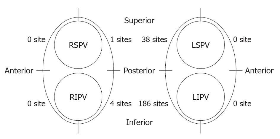
Figure 3 Position of sites where luminal esophageal temperature reached the cut-off temperature.
Most of the sites were located along the posterior side of the LPV, especially around the left inferior pulmonary vein (LIPV), while only five sites were observed near the RPVs. LSPV: Left superior pulmonary vein; RSPV: Right superior pulmonary vein; RIPV: Right inferior pulmonary vein.
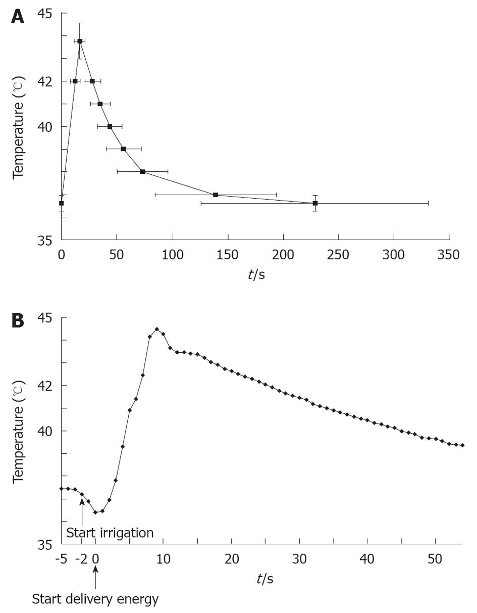
Figure 4 Luminal esophageal temperature.
A: Graph of luminal esophageal temperature (LET); B:Transient drop in the LET. After having caused a transient drop in the luminal esophageal temperature just before delivering energy, the luminal esophageal temperature reached the cut-off temperature.
- Citation: Sato D, Teramoto K, Kitajima H, Nishina N, Kida Y, Mani H, Esato M, Chun YH, Iwasaka T. Measuring luminal esophageal temperature during pulmonary vein isolation of atrial fibrillation. World J Cardiol 2012; 4(5): 188-194
- URL: https://www.wjgnet.com/1949-8462/full/v4/i5/188.htm
- DOI: https://dx.doi.org/10.4330/wjc.v4.i5.188


