Copyright
©2012 Baishideng Publishing Group Co.
World J Cardiol. May 26, 2012; 4(5): 173-182
Published online May 26, 2012. doi: 10.4330/wjc.v4.i5.173
Published online May 26, 2012. doi: 10.4330/wjc.v4.i5.173
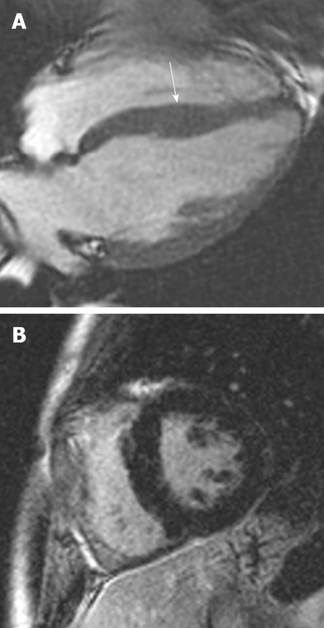
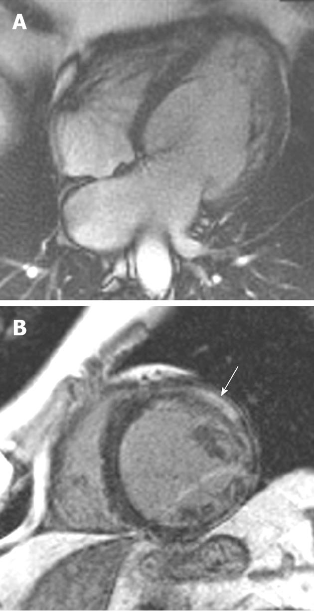
Figure 1 A 19-year-old man with Friedrich’s ataxia who presented with sudden onset chest pain.
A: Horizontal long-axis SSFP sequence showing circumferential hypertrophy (arrow, 14 mm thickness) of the anterobasal segment of the left ventricular; B: Late-enhanced sequence showing absence of high signal, characteristic of the hypertrophy seen in this disorder.
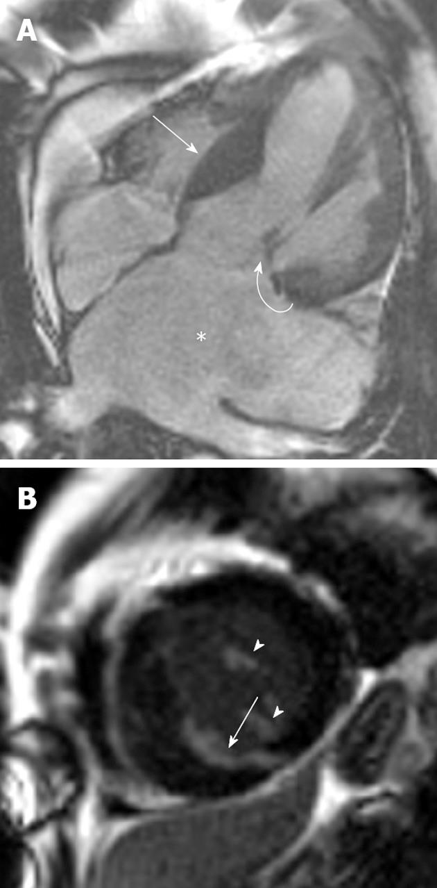
Figure 2 A 46-year-old man with Noonan’s syndrome.
A: Horizontal long-axis SSFP sequence showing septal hypertrophy (curved arrow) and a severely dilated left atrium secondary to severe mitral regurgitation (asterisk); B: Late-enhanced short axis sequence showing extensive late gadolinium enhancement (LGE) throughout the antero- and infero-septal myocardial segments (arrow). LGE was also noted in the papillary muscles (arrowheads).
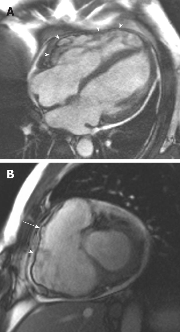
Figure 3 A 38-year-old man of Italian origin presented with palpitations and progressive shortness of breath during exercise.
A: Horizontal long-axis SSFP shows a heavily trabeculated right ventricle with thickened trabeculae and increased right ventricular (RV) volume. The RV free wall is very thin, with multiple small aneurysms (arrowheads); B: Short-axis SSFP sequence showing further small aneurysms of the RV free wall. Note again the increased RV volume.
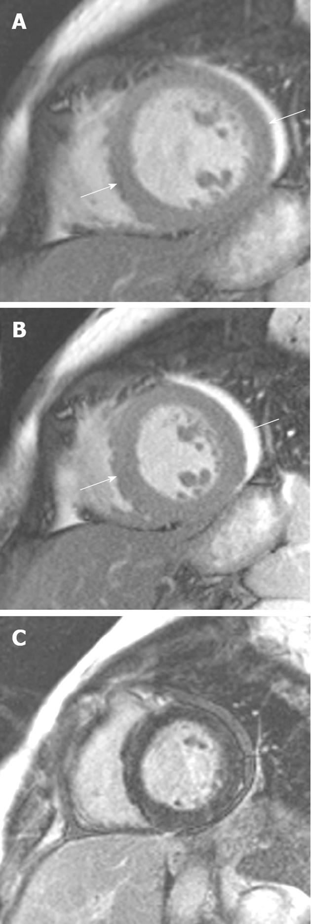
Figure 4 A 34-year-old woman 1 wk post-partum who developed progressive shortness of breath.
A: Short-axis SSFP sequence showing dilation of the left ventricle (end-diastolic diameter = 62 mm); B: The systolic phase demonstrating moderate circumferential hypokinesis (left ventricular ejection fraction 45%); C: Late gadolinium enhancement sequence showing an absence of high signal in this case.
Figure 5 A 46-year-old man with progressive heart failure and Becker’s muscular dystrophy.
A: Short-axis SSFP sequence showing moderate dilation of the left ventricle; B: Late gadolinium enhancement (LGE) sequence showing extensive transmural LGE throughout the lateral segments (arrow).
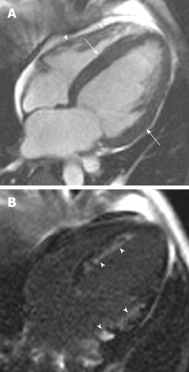
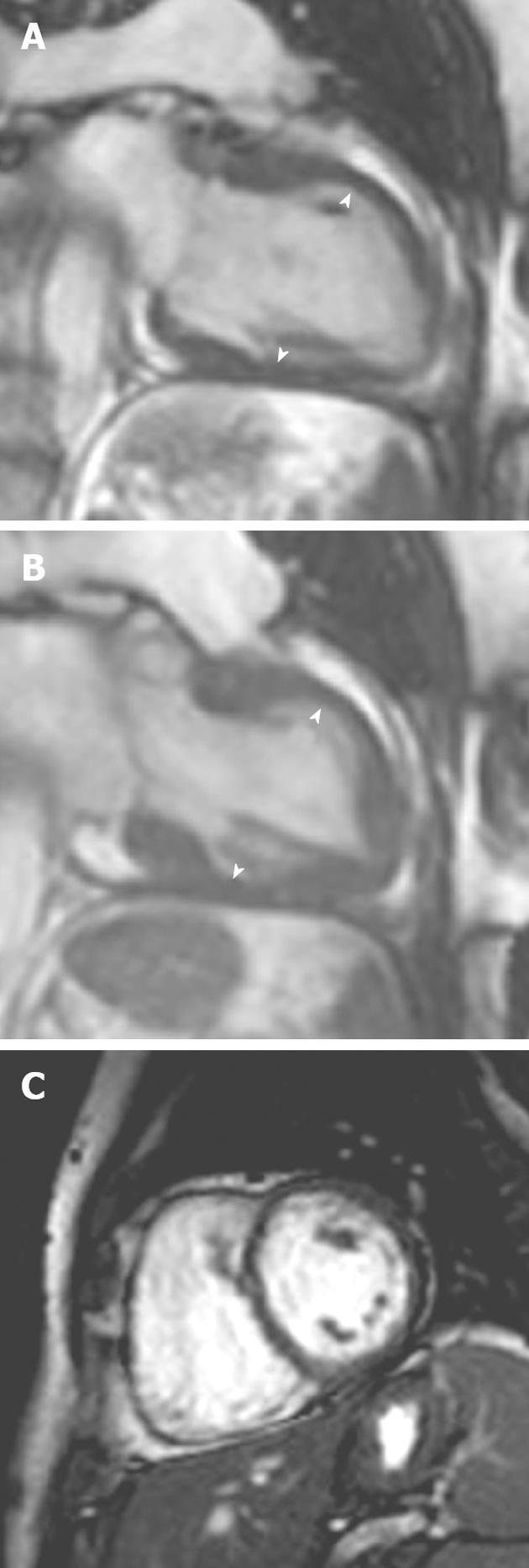
Figure 6 A 38-year-old man presented with progressive shortness of breath.
Serum measurements demonstrated hypereosinophilia. A: Horizontal long-axis SSFP sequence showing mild circumferential hypertrophy of the left ventricle (arrows) Note the normal thin right ventricular free wall (arrowheads), a useful differentiating feature from cardiac amyloid; B: Late-enhanced horizontal long-axis sequence showing extensive high signal in the typical subendocardial distribution (arrowheads) of eosinophilic myocardial infiltration. Cardiac amyloid is the principal other cardiomyopathy that mimics this late gadolinium enhancement appearance.
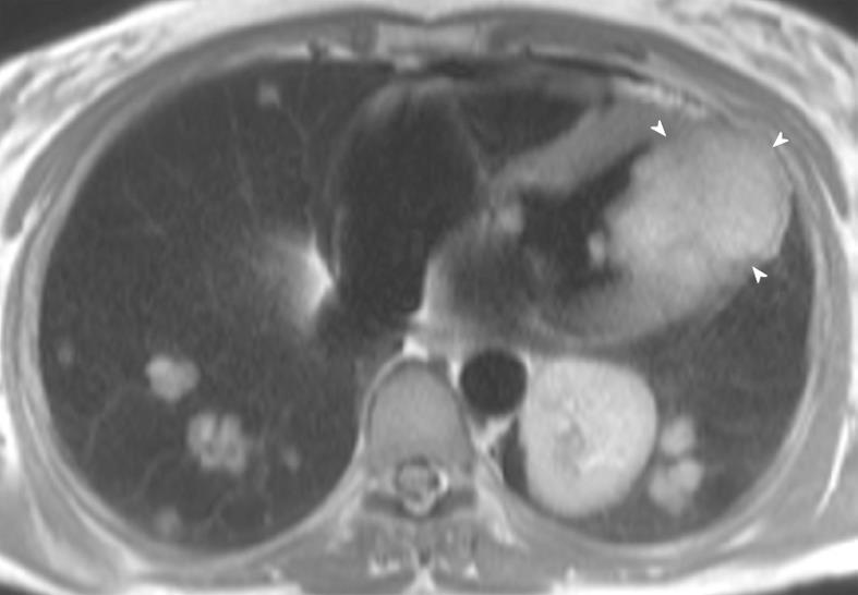
Figure 7 A 56-year-old woman with metastatic sarcoma.
Note the diffuse metastatic infiltration of the left ventricle (arrowheads), resulting in restrictive pathophysiology. There are also diffuse pulmonary metastases throughout both lungs.
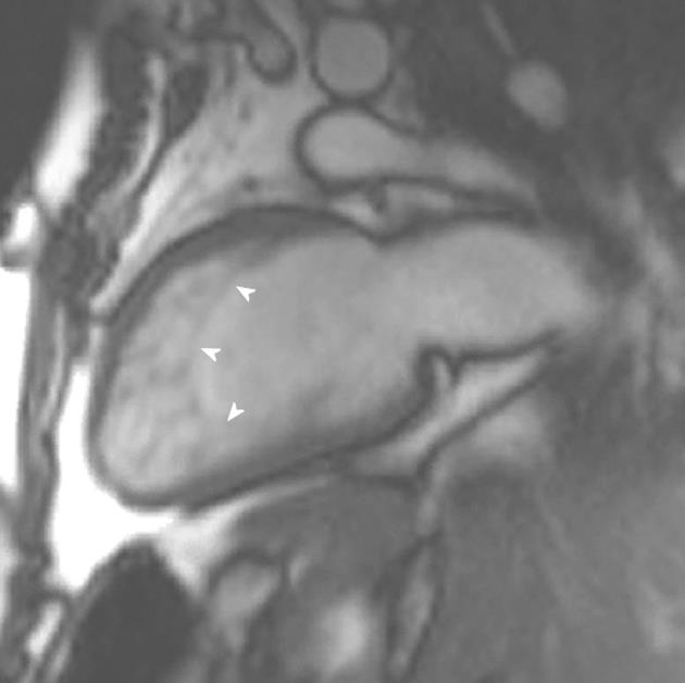
Figure 8 A 23-year-old man with Rubenstein-Taybi syndrome who presented with increasing shortness of breath.
Vertical long-axis view demonstrating increased trabeculations at the apical ventricular level fulfilling cardiac magnetic resonance imaging criteria for left ventricular noncompaction.
Figure 9 A 54-year-old woman with acute onset chest pain and palpitations following a road traffic accident.
A, B: Vertical long-axis SSFP sequence in (A) diastole and (B) systole showed akinesis of the left ventricular myocardial segments at the midventricular level; C: Late gadolinium enhancement showing no myocardial enhancement in the involved segments. Subsequent echocardiography 1 mo later showed normal mid-wall contraction.
- Citation: O’Neill AC, McDermott S, Ridge CA, Keane D, Dodd JD. Investigation of cardiomyopathy using cardiac magnetic resonance imaging part 2: Rare phenotypes. World J Cardiol 2012; 4(5): 173-182
- URL: https://www.wjgnet.com/1949-8462/full/v4/i5/173.htm
- DOI: https://dx.doi.org/10.4330/wjc.v4.i5.173


