Copyright
©2012 Baishideng Publishing Group Co.
World J Cardiol. May 26, 2012; 4(5): 148-156
Published online May 26, 2012. doi: 10.4330/wjc.v4.i5.148
Published online May 26, 2012. doi: 10.4330/wjc.v4.i5.148
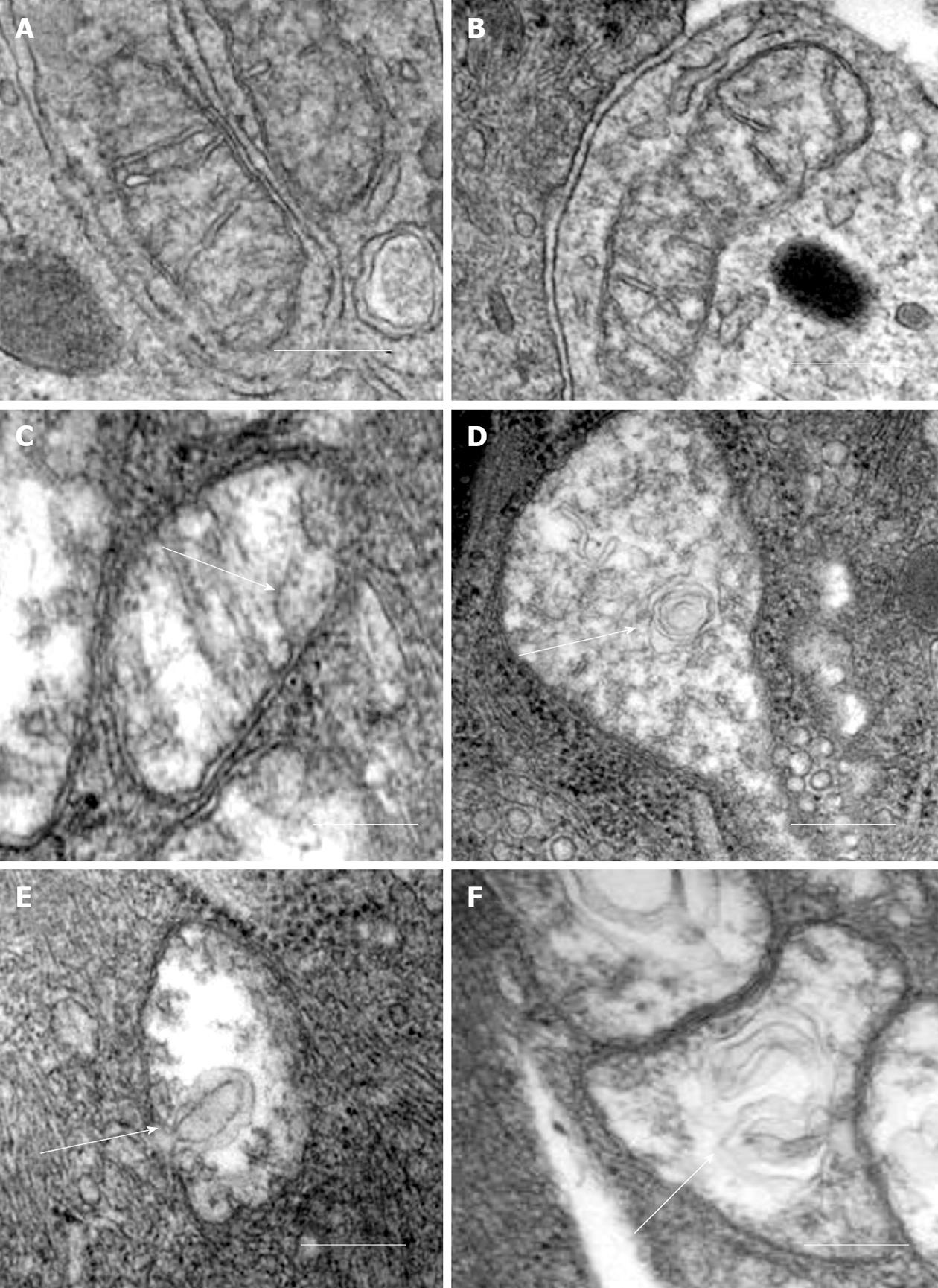
Figure 1 Different ultrastructural appearances of mitochondria in the arterial intima.
A, B: “Intact” appearances of mitochondria with well-defined cristae and well-preserved surrounding membranes; C-F: Destructive alterations of cristae and the formation of vacuole-like structures (shown by arrows) in zones of edematous matrix of mitochondria. Electron microscopy, scale = 200 nm.
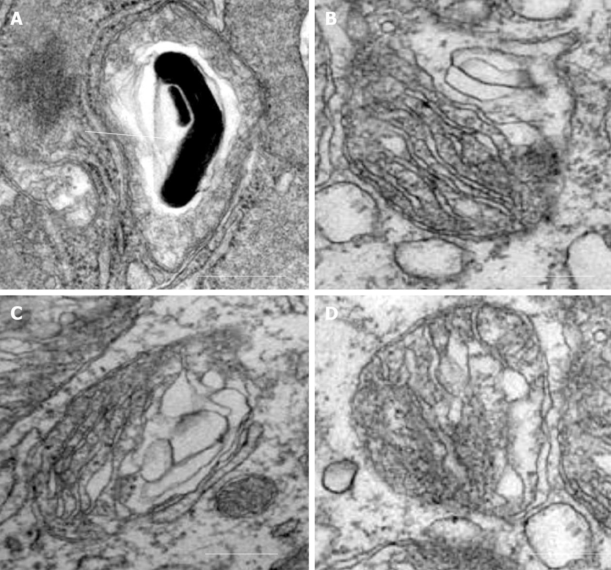
Figure 2 Alterations of mitochondria in intimal cells in atherosclerotic lesions.
A: Myelin-like structure within a swollen mitochondrion (shown by arrow); B-D: Destruction of cristae and surrounding membranes of mitochondria. Electron microscopy, scale = 200 nm.
- Citation: Chistiakov DA, Sobenin IA, Bobryshev YV, Orekhov AN. Mitochondrial dysfunction and mitochondrial DNA mutations in atherosclerotic complications in diabetes. World J Cardiol 2012; 4(5): 148-156
- URL: https://www.wjgnet.com/1949-8462/full/v4/i5/148.htm
- DOI: https://dx.doi.org/10.4330/wjc.v4.i5.148


